Clear Sky Science · en
A Multimodal Dataset to Investigate Task-Evoked Negative BOLD Response and Neurodegeneration
Why this brain study matters for everyday life
As we age, some people stay mentally sharp while others slowly lose memory and thinking skills. Doctors can see hints of these changes in brain scans, but one key signal, a drop in activity called the negative BOLD response, has been poorly understood. This article introduces a rich new collection of brain and health data from hundreds of adults that is designed to reveal what this “quieting” of brain regions really means for healthy aging and for conditions such as Alzheimer’s disease.
A closer look at the resting and working brain
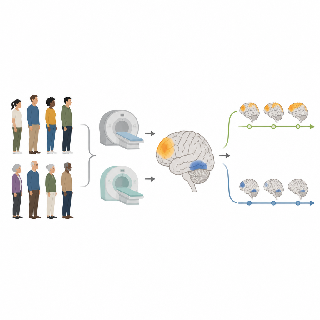
Modern brain scanners do more than take pictures of brain structure. They can also track tiny shifts in blood oxygen as we think, remember, and pay attention. Most studies focus on areas that “light up” during a task, but many regions instead turn down their activity. This quieting pattern helped scientists discover the brain’s default mode network, a set of regions that are active when our minds wander and less active when we concentrate. The new Quantitative Neuroimaging Laboratory Dataset was built specifically to study this lesser known signal by combining multiple types of scans and detailed thinking tests in both younger and older adults.
What the new dataset contains
The project enrolled 356 volunteers, including adults in their 20s and 30s and elders in their 60s and 70s, carefully screened to rule out dementia and serious medical problems. Each participant could take part in up to three long visits that blended brain imaging with paper and computer tests. The imaging side includes high resolution MRI to capture brain anatomy, specialized scans of blood flow and wiring, and both resting and task based functional MRI. In addition, three kinds of PET scans track brain energy use, amyloid plaques, and tau tangles, two hallmark proteins tied to Alzheimer’s disease. Many volunteers also gave blood for genetic testing and future analyses of blood based markers.
How thinking skills were tested
Inside the scanner, volunteers completed twelve different tasks that tap four broad abilities: solving new problems, remembering stories and word lists, recognizing vocabulary, and working quickly with symbols and patterns. Each task was carefully rehearsed beforehand so that performance during scanning reflected real thinking, not confusion about the rules. Outside the scanner, participants tackled a broad neuropsychology battery, from classic memory lists and story recall to puzzles of attention, language, and planning. Together, these measures show that younger adults generally respond faster and more accurately, while older adults often excel on knowledge based tasks like vocabulary, mirroring everyday experience.
How the brain data were cleaned and checked
Brain imaging is notoriously sensitive to tiny head movements and scanner quirks, so the team built an in house processing pipeline to standardize and clean every scan. Functional MRI data were aligned, corrected for the timing of slice acquisition, smoothed, and then passed through an automated method that strips out motion related noise. Additional steps removed leftover spikes from sudden shifts and filtered the signals to focus on meaningful slow rhythms. For PET scans, the researchers used automated tools to align the images with each person’s anatomy and to compute simple summary measures of how much tracer collected in each brain region, adjusting for non specific signal.
What early checks reveal about brain aging
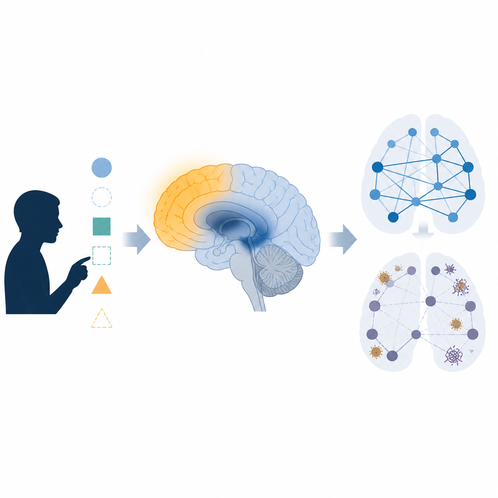
To confirm that the dataset behaves as expected, the authors compared groups using well known markers. In scans of sugar use, older adults showed lower brain metabolism than younger adults, matching decades of prior work. Amyloid and tau PET scans revealed that a sizable minority of cognitively normal elders already carry substantial deposits of these proteins, while younger adults have very little. Functional connectivity maps showed strong, distinct brain networks in both age groups and robust “see saw” patterns between networks that activate during tasks and those that deactivate. Task based scans confirmed that visual and movement regions turn on during challenges, while default mode regions turn down, especially in younger adults.
What this means for future brain health research
By bringing together many kinds of brain scans, genetics, blood samples, and detailed cognitive testing in the same people, this dataset offers a powerful way to study how patterns of brain deactivation relate to normal aging and early disease. Because the data are shared publicly in a standard format, other scientists can test ideas about how negative BOLD responses, brain networks, and disease related proteins interact over time. In the long run, a clearer picture of these links may help doctors read brain scans in a more personalized way, spotting who is aging normally and who may be on a path toward neurodegeneration long before symptoms become obvious.
Citation: Ghaderi Yazdi, B., Ozoria, S., Hojjati, S.H. et al. A Multimodal Dataset to Investigate Task-Evoked Negative BOLD Response and Neurodegeneration. Sci Data 13, 744 (2026). https://doi.org/10.1038/s41597-026-07081-x
Keywords: brain imaging, negative BOLD, functional MRI, Alzheimer biomarkers, cognitive aging