Clear Sky Science · en
“Dense Amygdala”: Extensive Complex-valued Functional MRI of the Ventral and Medial Temporal Lobe during Passive Movie Watching in Three Individuals
Why movies in the scanner matter
Most of us know movies can make us laugh, cry, or sit on the edge of our seats. This study asks what happens deep inside the brain during those rich emotional moments. By collecting an unusually large amount of brain data from just three people as they quietly watched full-length films, the researchers created a detailed resource for exploring how a key emotion hub, the amygdala, reacts to the sights and sounds of everyday social life on screen.
A close look at an emotion hub
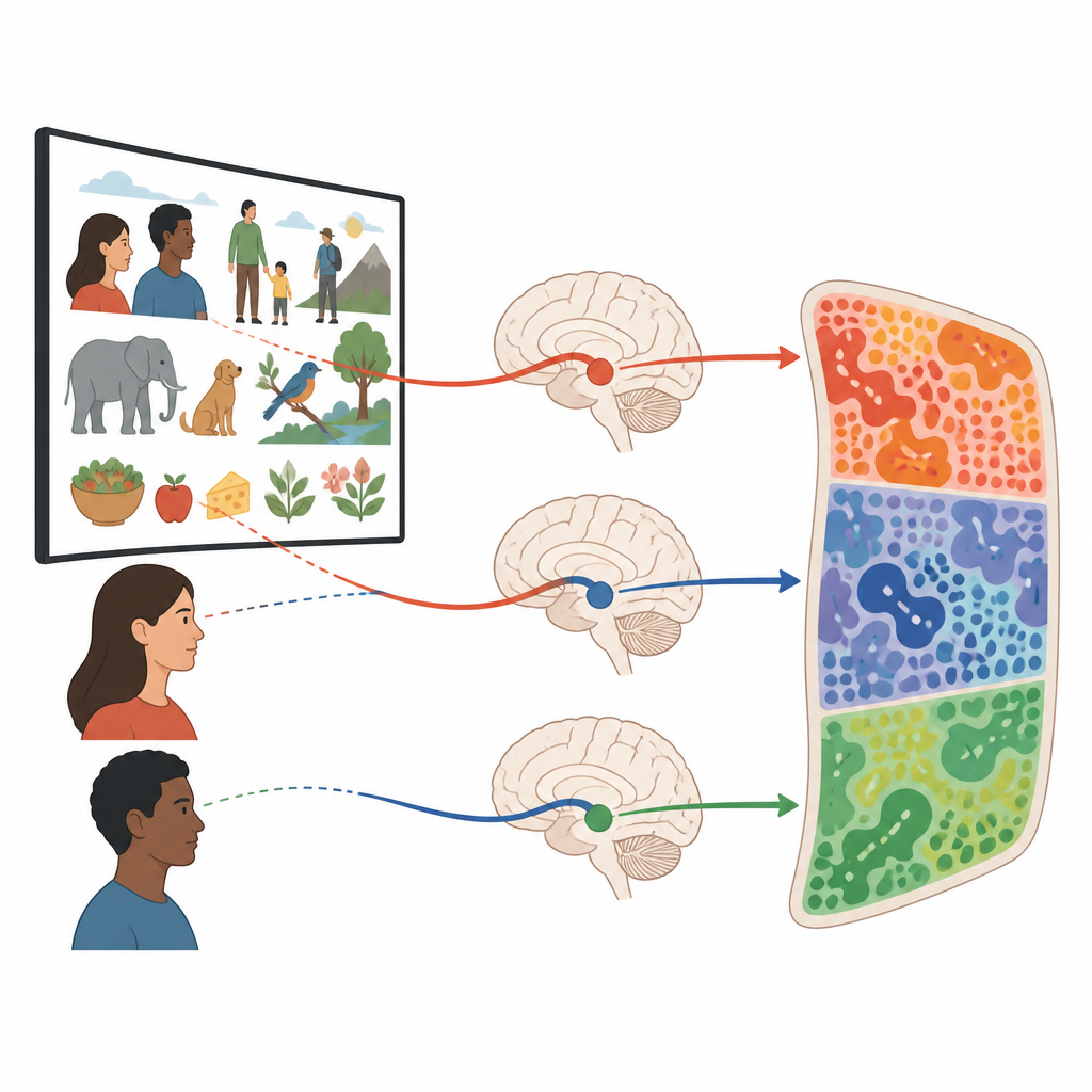
The amygdala sits deep in the temporal lobe and has long been linked to fear, reward, and the importance we attach to what we see and hear. Yet it is hard to study with standard brain scans, which often focus on the outer surface of the brain and average across many people. This project turns that logic around. Instead of scanning hundreds of volunteers briefly, the team scanned three adults for more than ten hours each while they watched four feature films and a fast-paced trailer montage. The scan setup was tuned to collect strong signals from the amygdala and nearby regions that support memory, vision, and social understanding.
Movies as natural stories for the brain
The film choices were not random. Two movies, Forrest Gump and The Grand Budapest Hotel, have been used in previous brain studies, making it easier to compare results across labs. Planet Earth episodes added scenes of animals and nature, while Jiro Dreams of Sushi focused on food and craft, themes thought to strongly engage the amygdala. The trailer mix packed intense social and emotional content into just a few minutes. Across twelve sessions, each participant saw movie segments, repeats of short clips, and a separate task with blocks of faces, objects, buildings, and scrambled images. After each session, they filled out mood and stress questionnaires, and later they rated how positive, negative, exciting, or anxious they felt every thirty seconds while rewatching the clips.
How the brain signals were captured and cleaned
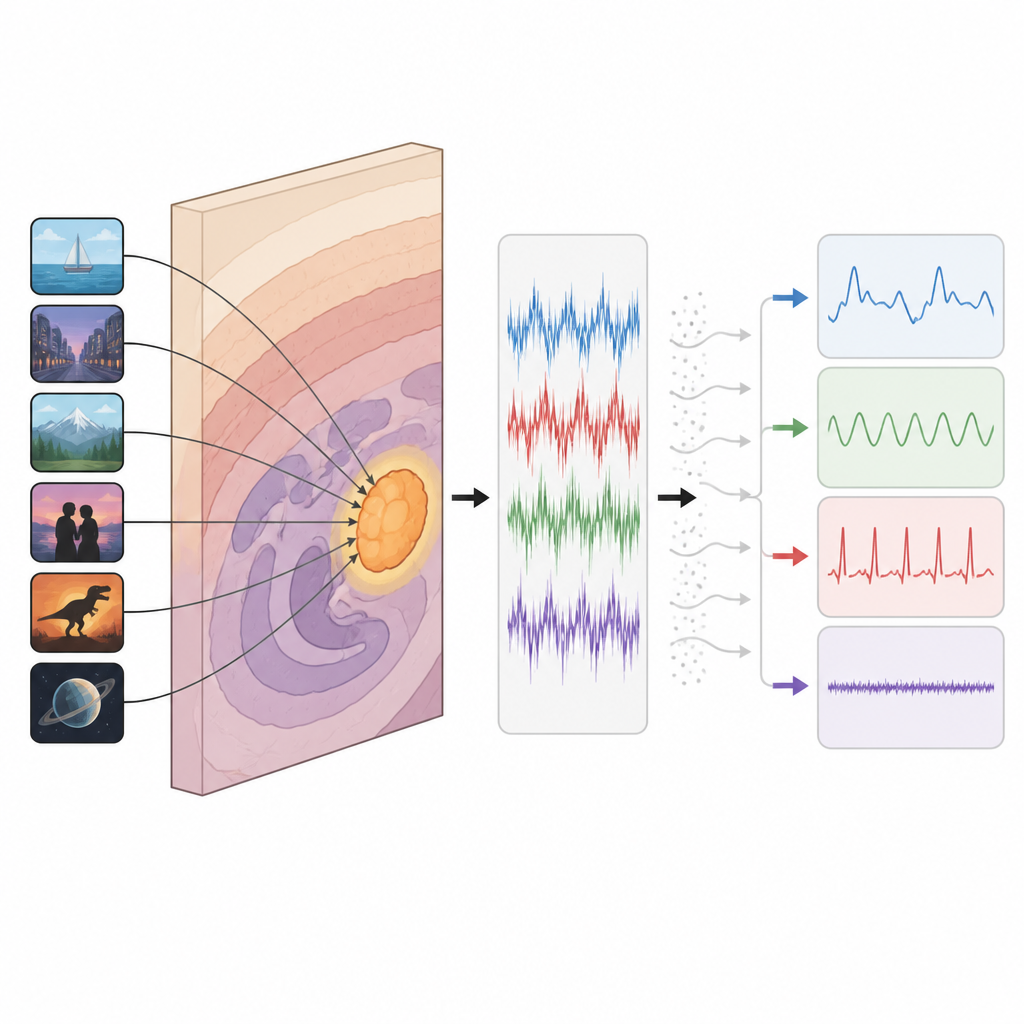
The researchers used a rapid type of MRI scan that samples brain activity roughly twice per second and keeps both the strength and phase of the signal. Focusing on a slab of tissue around the ventral and medial temporal lobes boosted sensitivity where it is usually weakest. Alongside the movie scans, they recorded heart activity, breathing, and pupil size to track arousal and body rhythms. A custom processing pipeline aligned all data to high-quality anatomical templates for each person and estimated how much of the signal was true brain response versus noise from head motion, scanner quirks, or blood vessels. Advanced math methods, including independent component analysis, split the data into patterns linked to respiration, heartbeat, and likely neurovascular responses tied to the movies.
What the dataset reveals so far
Initial checks show that the movies reliably evoked structured activity throughout the scanned brain slab, including the amygdala. When the team compared brain responses to faces in the movies with responses from the classic face-localizer task, they found similar patches in ventral temporal cortex, yet with clear differences between people. Measures of signal quality and head motion suggest that the data are stable enough to study fine-grained patterns. At the same time, the three volunteers differ in how strongly their amygdala appears in these patterns and in psychological traits such as positive affect, highlighting the very individuality this project aims to capture rather than smooth away.
How others can use this resource
All brain images, physiological recordings, low-level video and audio features, and emotion ratings are openly shared in a standard format. Researchers can use them to test new ways of cleaning brain data, to model how specific movie moments drive amygdala and ventral visual activity, or to compare complex-valued and traditional MRI analyses. Because the sample is tiny but densely studied, it is not meant to represent the general population. Instead, it offers a high-resolution case study of three brains, helping scientists design smarter future experiments on social and emotional processing, and deepening our understanding of how the amygdala responds when stories on a screen feel vivid and real.
Citation: Tyszka, J.M., Diamandis, Z., Keles, U. et al. “Dense Amygdala”: Extensive Complex-valued Functional MRI of the Ventral and Medial Temporal Lobe during Passive Movie Watching in Three Individuals. Sci Data 13, 738 (2026). https://doi.org/10.1038/s41597-026-07065-x
Keywords: movie fMRI, amygdala, social neuroscience, naturalistic stimuli, brain imaging data