Clear Sky Science · en
A multimodal biomechanics dataset with synchronized kinematics and internal tissue motions during reaching
Why the hidden motion inside muscles matters
Every time you reach for a mug or hand someone an object, your arm seems to move as a single, solid piece. In reality, it is a layered, sliding bundle of muscles, fat, skin, and bone, all shifting in complex ways beneath the surface. These hidden motions may hold clues to why some people move with effortless precision, why others develop pain or injury, and how to design better therapies, sports training, and even robots. This study introduces a rich public dataset that lets researchers, clinicians, and engineers look under the skin while people perform a simple reaching task, linking what the arm does on the outside to what tissues do on the inside.
Looking beneath the surface of a simple reach
The researchers focused on a very basic movement: slow, rhythmic forward reaches made while standing, with the hand moving from the hip to in front of the body and back again. Thirty-six healthy adults took part, spanning three broad skill levels: world‑class performers and athletes, region‑level or long‑term recreational movers, and people with no formal movement training. By choosing a simple, goal‑free task and pacing it with a visual metronome, the team minimized the effects of reaction time, strategy, and sport‑specific tricks. Instead, the design was meant to expose more general features of how people actually control their muscles and manage subtle tremors when moving slowly, which is a situation humans typically find surprisingly difficult.
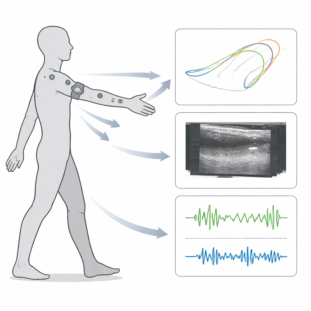
Many sensors watching the same motion
To capture both the visible arm motion and the hidden internal activity, the study combined several types of sensors that recorded at the same time. External movement of the shoulder, upper arm, elbow, forearm, and hand was tracked in 3D using reflective markers and a ring of high‑speed cameras. A set of small wireless devices on the biceps, triceps, and palm measured muscle electrical activity and tiny accelerations, which are useful for detecting tremors. Most unusually, a slim ultrasound probe was strapped around the upper arm to image a cross‑section of the triceps and brachialis muscles, along with the surrounding fat and the humerus bone. This created tens of thousands of “movie frames” of the inside of the arm as it moved.
Turning raw recordings into usable signals
Collecting all these data streams is only half the challenge; they also need to be precisely lined up in time and cleaned before others can use them. Each sensor system runs on its own clock, so the team used electronic timing pulses and careful analysis to calibrate how the clocks drifted relative to one another, then mathematically rescaled and shifted the data so that, for example, a burst of tremor in the accelerometer aligns with the matching wiggle in joint motion and tissue movement. Motion capture traces were filtered and reduced to a single main “arm movement” signal that made it easy to detect each reach cycle. Muscle signals were filtered and converted into smooth envelopes showing how strongly each muscle was active. Tremors were isolated by focusing on a specific frequency band in the accelerometers, and automatic algorithms were used—then checked by eye—to mark when tremors started and stopped.
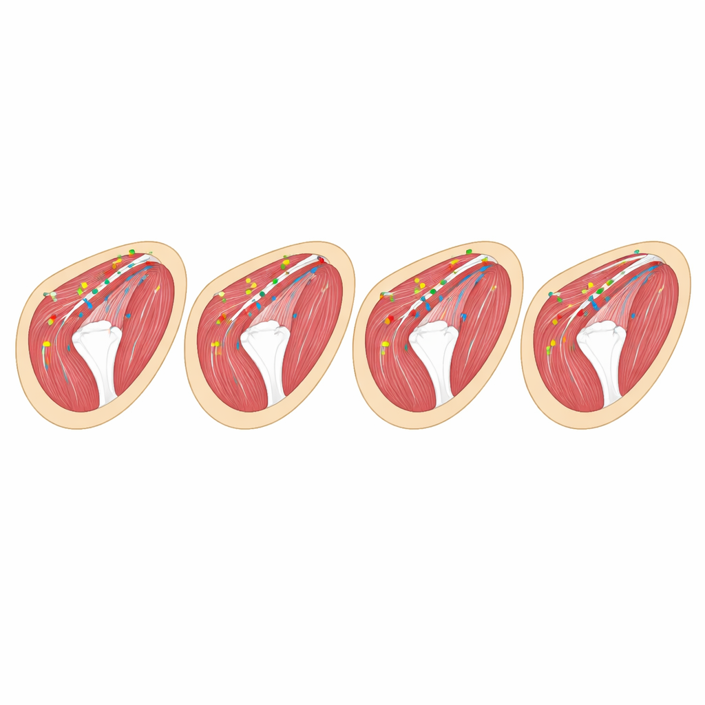
Following individual points inside muscle
The standout feature of this dataset is that it does not just store ultrasound images; it also includes the tracked paths of 11 specific points inside each video, followed frame by frame across about 300,000 images. Some of these points sit on the humerus bone and on the boundary between two muscles, while others lie within different zones of the triceps and brachialis. To create these tracks, the authors used a semi‑automated workflow that starts with humans labeling a modest number of frames, then trains a deep‑learning model to follow the same points through the full movie, and finally applies optical‑flow tools and manual corrections where needed. The result is a dense, continuous description of how tiny regions of tissue slide, stretch, and deform relative to one another during reaching—information that is nearly impossible to obtain non‑invasively by other means.
A shared resource for movement, health, and AI
All recordings, derived measures, and code are freely shared in standard formats, along with tutorials for loading and visualizing the data in common scientific software. Because the dataset links joint‑level motion, muscle activity, tremors, and internal tissue movement across people with different skill levels, it can be used to explore how expert movers differ from novices, how forces travel through the arm, or how subtle tissue behavior might relate to pain or performance. At the same time, the frame‑by‑frame ultrasound point tracks provide a rare training ground for modern deep‑learning systems that aim to automatically follow structures in medical images. In short, this work does not answer a single narrow question; instead, it offers a carefully validated foundation on which many future studies of human movement and medical image analysis can be built.
Citation: Pallarès-López, R., Folgado, D., Magana-Salgado, U. et al. A multimodal biomechanics dataset with synchronized kinematics and internal tissue motions during reaching. Sci Data 13, 709 (2026). https://doi.org/10.1038/s41597-026-07019-3
Keywords: human movement, ultrasound imaging, muscle mechanics, biomechanics dataset, deep learning tracking