Clear Sky Science · en
Image-to-molecule benchmarking dataset with fractal pattern and hierarchical morphology recognition
Why Tiny Crystal Patterns Matter
When a drop of a chemical solution dries, it can leave behind surprisingly beautiful and intricate crystal landscapes. This paper explores how those patterns are not just pretty pictures: they quietly encode information about the molecules themselves. The authors present a large, open image collection that links the shapes seen under microscopes to the underlying chemistry, creating a playground for artificial intelligence to learn how a molecule’s structure shows up in its visible form.
Pictures That Reveal Hidden Structure
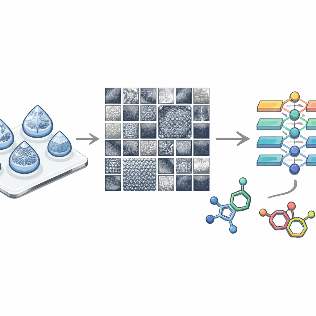
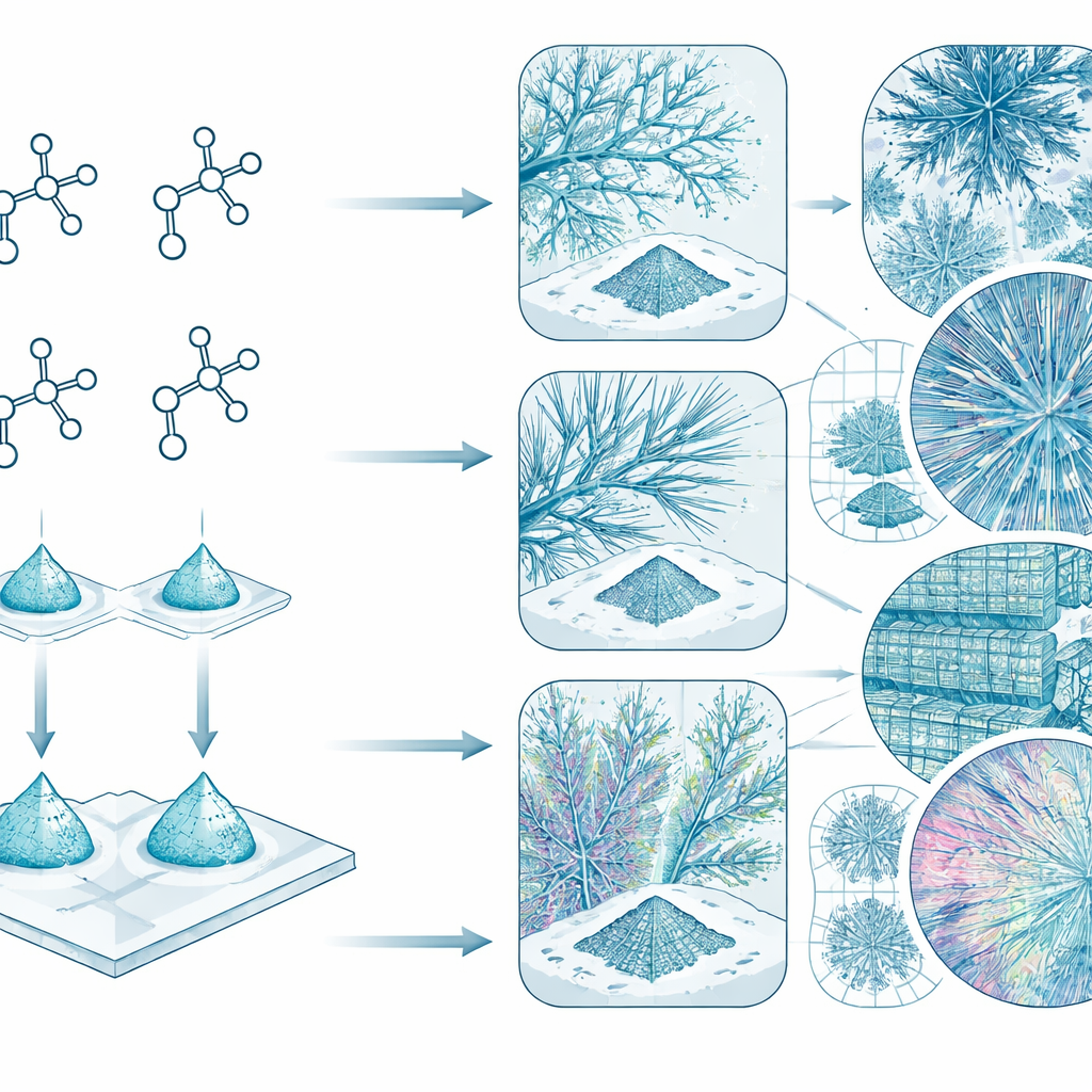
The study focuses on a family of related compounds called quaternary phosphonium salts. These materials are solid at room temperature and can form crystals with strikingly different appearances, even when their molecules differ by only a single small fragment. Using scanning electron microscopes and optical microscopes, the team recorded more than 3,500 high‑resolution electron images and nearly 400 optical images from 19 such compounds and 10 of their mixtures. Each image captures the way crystals grow, branch and organize themselves as droplets of solution dry on a surface.
A Library of Shapes Across Many Scales
The researchers designed the image collection so that the same types of structures could be compared fairly. For each compound, they took at least 100 electron microscope images at 14 carefully chosen magnifications, from broad overviews of an entire dried droplet down to fine details just tens of nanometers across. Additional images of mixtures were taken at many “in‑between” magnifications to test how well computer models can handle new, slightly different viewing conditions. Optical microscope images, taken at lower magnification, echo the same patterns and can be used alongside electron images for more creative image‑based methods.
Fractals, Layers, and Crystal Landscapes
One of the most eye‑catching findings is the extraordinary variety of shapes. Some compounds form clearly faceted crystals with sharp edges, while others give smoother, melted‑looking deposits. Within a single compound, several distinct micro‑landscapes can appear, hinting at different crystal forms. Common motifs include tree‑like, branching “fractal” structures, needle bundles, grid‑like lamellae, and complex layered textures. These patterns repeat in a hierarchical way: large structures are built from smaller, similar elements, which can still be recognized when the image is zoomed in or out, much like looking at a coastline from different altitudes.
From Images to Molecules and Back Again
Crucially, earlier work by the authors showed that a deep‑learning model can already tell apart closely related members of this compound family using only microscopy images. That result implies that the visual appearance of the crystals truly reflects subtle differences in molecular structure. The newly published dataset goes further by making the full, curated image collection public, complete with imaging settings and organized folders. This opens the door to two complementary lines of machine‑learning research: algorithms that read a microscopy image and infer what kind of molecule produced it, and algorithms that take a molecular description and generate plausible crystal patterns that might be seen in the lab.
What This Means for Future Materials
For non‑specialists, the big takeaway is that the shapes seen under a microscope are not random; they are fingerprints of the molecules that formed them. By pairing thousands of carefully documented images with known chemical structures, this work creates a benchmark resource for researchers who want to teach computers to understand and even design new materials based on their appearance. In time, such tools could help chemists quickly screen compounds, optimize manufacturing steps, or deliberately engineer crystal patterns that give materials better performance in technologies ranging from electronics to medical drugs.
Citation: Arkhipova, D.M., Boiko, D.A., Oganov, A.A. et al. Image-to-molecule benchmarking dataset with fractal pattern and hierarchical morphology recognition. Sci Data 13, 570 (2026). https://doi.org/10.1038/s41597-026-06941-w
Keywords: microscopy images, materials morphology, machine learning, crystal patterns, materials discovery