Clear Sky Science · en
A spatial transcriptomics comparison of the adult versus metamorphosed axolotl brain
Why a salamander’s brain matters to us
Imagine an animal that can regrow parts of its brain after injury and stays healthy far longer than you might expect. The Mexican axolotl is just such a creature. Unlike most vertebrates, axolotls can repair complex body parts, including parts of the central nervous system. But when these animals are pushed to leave their youthful, water-dwelling form and become land-dwellers, they steadily lose much of this repair power. This study maps, in fine detail, how the cells and genes in the axolotl brain are arranged before and after this life change, creating a reference atlas that could ultimately help researchers understand—and perhaps one day boost—regeneration in other animals, including humans.
A shape-shifting animal with unusual healing powers
Axolotls are famous for staying in a “teenage” aquatic state even after they are able to reproduce, keeping traits like feathery external gills. In this state, they can regrow limbs, parts of the eye, the spinal cord, and even portions of the brain. Under certain conditions, such as exposure to thyroid hormone, adult axolotls can be forced to undergo metamorphosis, losing their gills and adopting a more typical salamander body suited to life on land. This shift, however, comes with a cost: their ability to regenerate declines, and their lifespan shortens. Until now, scientists have lacked a brain-wide, cell-by-cell view of what changes inside the head of an axolotl as it makes this transition.
Reading the brain as a map of cells and molecules
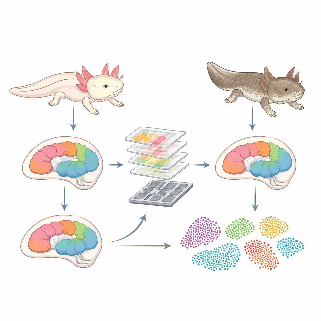
To fill this gap, the researchers used a technique called spatial transcriptomics, which allows them to see which genes are active in individual cells, while preserving each cell’s position in the tissue. They applied a high-resolution version of this method, called Stereo-seq, to brain slices from five major regions: the olfactory bulb, telencephalon, diencephalon/mesencephalon, rhombencephalon, and pituitary. Brains from water-dwelling adults were compared with those from animals that had been driven to metamorphose. After careful preparation, imaging, and sequencing, the team ended up with over 83,000 high-quality cells, each tagged with its own gene activity profile and precise coordinates in the brain.
Who’s who in the axolotl brain
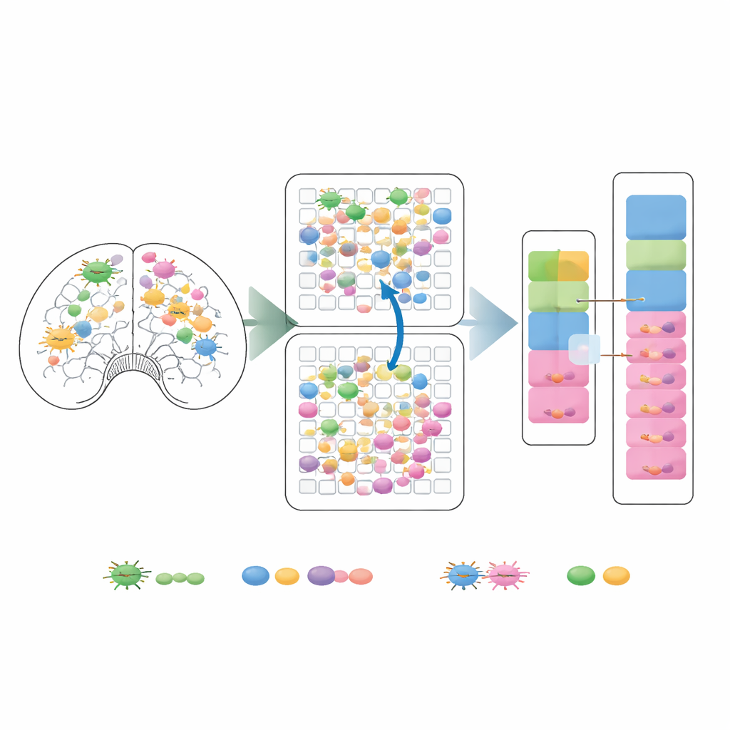
By clustering cells with similar gene activity, the team identified 24 distinct cell types spread across the brain. These included multiple kinds of neurons, support cells that wrap nerve fibers, blood vessel-associated cells, immune-like microglial cells, and hormone-producing cells in the pituitary. Of special interest were ependymoglial cells, which line brain cavities and are known to give rise to new neurons during repair. Earlier work had shown that some of these cells become activated during brain regeneration after injury. In this study, the authors found several subtypes of these cells and mapped exactly where they live in different brain regions, both before and after metamorphosis.
How metamorphosis reshapes brain cell communities
With this atlas in hand, the researchers asked how the “cellular cast” and gene activity patterns shift between the water-dwelling and metamorphosed brains. Overall, the major cell types and their spatial layouts remained broadly similar, showing that the basic brain architecture is preserved. Yet there were clear, focused changes. Certain ependymoglial subtypes, particularly one found near a brain region called the infundibulum, showed large numbers of genes turning up or down during metamorphosis, including genes linked to immune function and hormone signaling. At the same time, microglial cells became more abundant, and the inferred strength of communication between microglia and ependymoglial cells increased, hinting that immune-like signals may play a stronger role in the metamorphosed brain.
A shared resource for future regeneration research
This work does not attempt to fully explain why regeneration fades after metamorphosis, but it lays essential groundwork. The study delivers a well-validated, publicly available map of cell types, their locations, and their gene activity across key brain regions, before and after metamorphosis. For non-specialists, the takeaway is that losing regenerative capacity is not just a matter of whole organs changing shape; it is also about precise shifts in particular support and immune-related cells and how they talk to each other. By making these detailed data and analysis tools freely accessible, the authors provide a foundation for future experiments that can probe which of these cellular and molecular changes truly tip the balance between a brain that can rebuild itself and one that cannot.
Citation: Wang, S., Fu, S., Liu, X. et al. A spatial transcriptomics comparison of the adult versus metamorphosed axolotl brain. Sci Data 13, 509 (2026). https://doi.org/10.1038/s41597-026-06917-w
Keywords: axolotl brain, regeneration, metamorphosis, spatial transcriptomics, neural stem cells