Clear Sky Science · en
A dense longitudinal multimodal single-subject rs-fMRI dataset acquired by self-administered scanning
Why one person’s many brain scans matter
Imagine being able to watch a single brain over nearly a year, day after day, while its owner goes about normal life, changes medication, and moves from winter into summer. This article describes exactly that: an unusually rich set of brain scans and daily-life records collected from one researcher who learned to run the hospital MRI scanner on himself. The result is a public dataset that anyone can use to test new analysis methods, study how brain signals change over time, and teach the next generation of neuroscientists what real-world data look like.
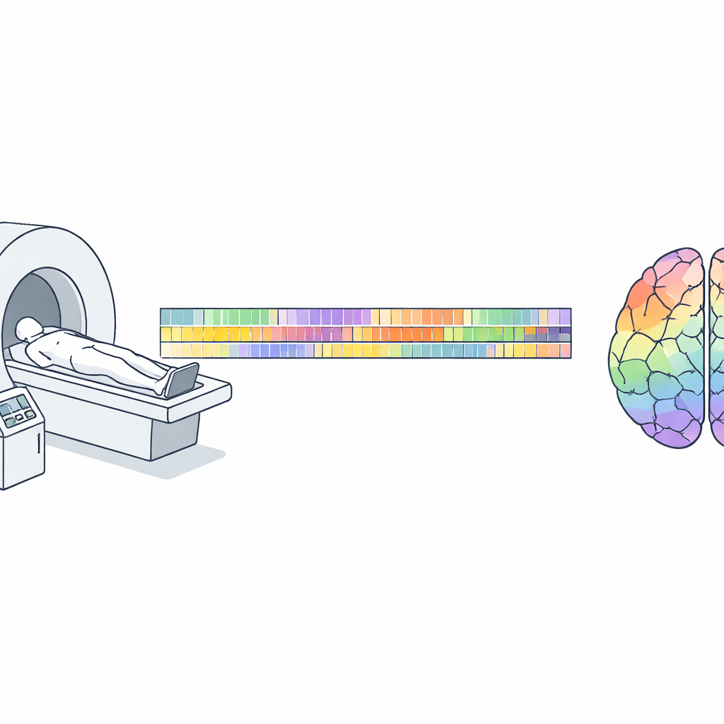
One dedicated volunteer, many hours in the scanner
The study followed a single 34-year-old researcher for 11 months, during which he lay in a clinical 3‑Tesla MRI scanner for a total of 85 hours of resting-state brain scans. About 52 of those hours came from a tightly standardized routine: in 128 sessions over 7.5 months, he first kept his eyes open for 10 minutes, then closed them for 14 minutes while the scanner measured spontaneous brain activity. Alongside this, he collected 195 high‑resolution pictures of brain structure and 54 sessions of diffusion scans that map the brain’s wiring. To put this in context, this one person’s dataset rivals, in depth over time, what large projects have achieved with far more resources—but using equipment and settings much closer to everyday clinical practice.
Self-scanning on a hospital machine
Most MRI studies rely on trained staff to position volunteers, start scans, and watch over safety. Here, after careful ethics review and safety training, the researcher did almost everything himself for the majority of sessions. He slid into the scanner, aligned his head using visible laser crosshairs aimed relative to his eyes, and launched preset scan sequences through the standard console. Early in the project, head position varied more from day to day; once the eye-based alignment method was adopted, the scans became remarkably consistent, with typical differences of less than 3 millimeters and about one degree between sessions—good enough for precise comparisons across months.
Watching motion, alertness, and sleep
Because even tiny movements can blur brain images, the study paid close attention to motion and wakefulness. Automated quality checks showed that 58 hours of functional data met a strict low-motion criterion, and more than 75 hours met a moderate one. When the participant was awake with eyes open, movement was minimal; with eyes closed or drifting toward sleep, motion rose in a predictable way, and full sleep produced the most motion—still within a useful range for some types of analysis. The researcher also recorded breathing and pulse during many runs, rated how sleepy he felt, and noted whether he dozed off, creating a rare combination of brain activity, body signals, and subjective state across the full spectrum from alertness to sleep.
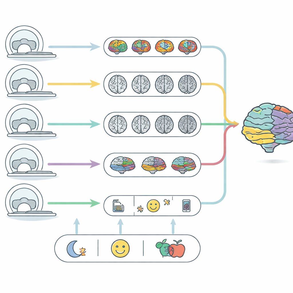
Daily mood, medication, and lifestyle in the picture
Beyond the scanner, the participant logged his antidepressant medication (venlafaxine) in fine detail as he tapered off over several months, including doses, timing, and even capsule bead counts. He tracked sleep times, coffee and alcohol use, exercise, and step counts through a private messaging system and phone sensors. Before most sessions he completed a brief reaction-time test to measure alertness and a standard mood questionnaire. From these raw records, the author computed simple summaries—such as recent dose, a rolling three‑week average, and indicators of possible withdrawal—that can be aligned with each scan. Crucially, the paper stresses that these overlapping changes in medication, season, and scanning skill all march along the same time line, making it impossible to say which factor causes any given brain change. They are meant as context, not proof of cause and effect.
What this resource is—and what it is not
All of the data are organized under a widely used standard for brain imaging studies and released into the public domain on the OpenNeuro platform, along with code used to clean and summarize them. This makes the dataset ideal for testing new preprocessing pipelines, comparing different quality-control strategies, studying how stable brain measures are within a single person, and teaching students how real datasets are structured. At the same time, the author is clear about its limits: it covers only one brain; some corrections routinely used in research (such as certain distortion fixes) are missing; and scanner drift cannot be separated from biological change. For a lay reader, the key takeaway is that a determined individual, working within careful safety and ethical boundaries, can turn a hospital scanner into a long-term personal observatory of the brain—providing a powerful sandbox for methods and education, rather than a final word on how drugs, seasons, or moods shape the mind.
Citation: Petrovskiy, E.D. A dense longitudinal multimodal single-subject rs-fMRI dataset acquired by self-administered scanning. Sci Data 13, 495 (2026). https://doi.org/10.1038/s41597-026-06879-z
Keywords: resting-state fMRI, longitudinal brain imaging, self-administered MRI, single-subject dataset, neuroimaging methods