Clear Sky Science · en
3D+t human sperm flagellum centerline dataset
Why the motion of tiny swimmers matters
When people talk about fertility, they often focus on hormone levels or sperm counts. But another crucial factor is how well sperm actually swim. Their whip-like tails, called flagella, power a long journey through the female reproductive tract, and subtle changes in this motion can mean the difference between success and failure. Until now, most research has seen this movement only as flat, two-dimensional shadows on a screen. This article presents a new three-dimensional dataset that finally captures how human sperm tails move in real space and time, opening the door to more precise fertility tests and smarter computer tools.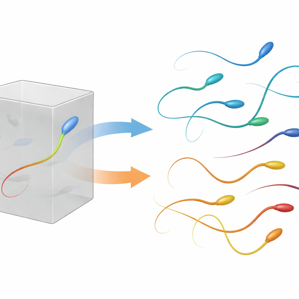
From flat movies to full 3D paths
For decades, standard laboratory systems have judged sperm by watching how their heads trace paths in two dimensions under a microscope. These systems calculate speeds and straightness from those tracks, but ignore how the tail itself beats and how the cell moves up and down in depth. In reality, sperm swim in a fully three-dimensional environment, and their tails follow complex, looping paths. Earlier image collections mostly offered still images or simple 2D movies, often of just the head or of sperm from other species, or of cells tethered to a surface instead of freely swimming. A few groups had begun to record 3D motion, but their data were hard to access or involved only a small number of cells.
Building a richer picture of sperm swimming
The authors introduce 3D-SpermFlagella, the first large, openly available collection dedicated to the 3D motion of human sperm tails. Using a custom high-speed microscope, they recorded stacks of images at many focal depths while the sample was rapidly moved up and down, effectively scanning a tiny 3D volume around each cell dozens of times per second. Each recording captures free-swimming sperm in a warm liquid environment close to body temperature, preserving their natural behavior. From these recordings, the team reconstructed the exact 3D curve traced out by each tail at every instant, from the neck where it joins the head to the thin tip at the far end.
How the tail lines were traced
Turning raw images into clean tail paths required a mix of human judgment and computer power. The researchers first used a graphical tool to click on the tail tip in 3D views, because this faint, narrow region is difficult for software to locate reliably. The brightest region of each image volume – the sperm head – served as the starting point. A specialized algorithm then searched for the least “costly” path through the image, favoring bright, tail-like structures between head and tip. Every resulting trace was checked visually from several angles; if the computed path wandered off the tail due to noise, sharp bends, or optical artifacts, the team adjusted the start or end points and re-ran the tracing. Finally, they converted pixel positions into real-world distances in micrometers and smoothed each curve so that points were spaced at regular intervals along the tail.
What the dataset contains
In total, the collection holds 135 human sperm cells, each tracked for about two seconds of swimming with a fine time step of one ninetieth of a second. For every time point, three files store the X, Y, and Z coordinates of hundreds of points along the tail, from neck to tip, either in pixel units or in physical units. The cells fall into two biologically important groups. Some were kept in a simple solution that does not trigger the changes needed for fertilization, known as non-capacitating conditions. Others were exposed to a medium that encourages the more vigorous behavior seen near the egg, called capacitation. This second group includes cells that display hyperactivation – a wilder, more forceful beat pattern thought to help sperm navigate thick fluids and escape surface traps.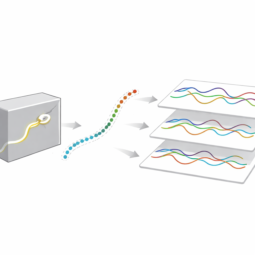
Why this matters for medicine and machines
By making these detailed 3D tail paths freely available, the authors provide a foundation for both biology and computer science. Researchers can now test theories of how sperm generate thrust, turn, or respond to their surroundings using real human data instead of idealized models or flat projections. Clinicians and engineers can compare classic two-dimensional motility scores with full 3D measures, potentially revealing hidden problems in sperm performance that current tests miss. At the same time, the dataset serves as a high-quality “answer key” for training and evaluating artificial intelligence systems that automatically find and track sperm tails in microscope images. In short, this work does not claim a new biological discovery about sperm by itself; instead, it delivers the precise, three-dimensional raw material that others will use to better understand, diagnose, and perhaps one day improve human fertility.
Citation: Hernández-Herrera, P., Hernández, H.O., Bribiesca-Sanchez, A. et al. 3D+t human sperm flagellum centerline dataset. Sci Data 13, 505 (2026). https://doi.org/10.1038/s41597-026-06876-2
Keywords: sperm motility, 3D microscopy, flagellum tracking, fertility research, bioimage analysis