Clear Sky Science · en
Multisession fNIRS-EEG data of Post-Stroke Motor Recovery. Recordings During Intact and Paretic Hand Movements
Why this matters for life after stroke
Stroke is one of the main causes of long-term disability, and many survivors struggle with basic arm and hand movements needed for everyday tasks. Doctors can see the damaged area on a brain scan, but they still have limited tools to track how the brain rewires itself during rehabilitation. This article presents a new open dataset that follows stroke patients over many therapy sessions, recording brain activity with two different non-invasive methods while they move both their affected and healthy hands. The resource is designed to help researchers build better rehabilitation strategies and future brain-controlled assistive devices.
Watching the healing brain in action
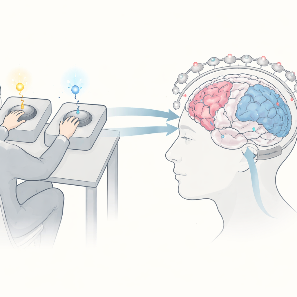
The study focuses on people with weakness in one arm after a stroke. Sixteen adults with moderate disability took part, most in the early months after their stroke, when the brain is thought to be especially adaptable. Over a two-week inpatient rehabilitation period, each person completed between three and six experimental sessions on separate days. During these sessions, they performed a simple reaction-time task: seated at a table with both hands in a custom box, they watched small lights above two buttons. When the light above the “target” hand flashed, they tried to press the matching button, while ignoring flashes on the other side. This setup allowed the researchers to compare brain activity during movements of the impaired (paretic) and intact hands.
Two gentle windows into brain activity
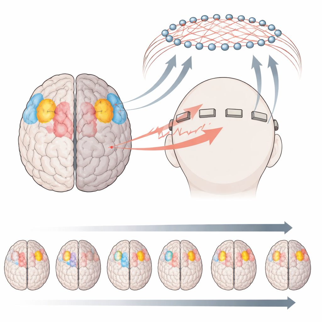
While patients performed the task, their brain signals were recorded in two ways. First, functional near-infrared spectroscopy (fNIRS) shined weak infrared light through the scalp to monitor changes in blood oxygen in the outer layers of the brain, especially over the regions that control movement. These changes reveal how hard a given area is working. The system used dozens of light sources and detectors across both sides of the head, forming 70 measurement channels. Second, electroencephalography (EEG) measured the brain’s fast electrical activity from eight electrodes interleaved among the fNIRS sensors, plus additional sensors for muscle activity and heart rhythm. A custom hardware trigger ensured that both systems were precisely time-locked to each light flash and button press, so researchers can align brain signals with behavior down to fractions of a second.
What the first checks on the data reveal
To show that the dataset is scientifically useful, the authors walked through example analyses from one patient with a stroke in the left side of the brain and weakness in the right hand. Using fNIRS, they filtered the signals and converted light changes into estimates of oxygenated and deoxygenated blood. Maps of blood flow over time showed an early response in the damaged (left) hemisphere when the weak hand moved, followed by strong activity in the opposite (right) hemisphere. This pattern suggests that the healthier side of the brain may be recruited to help compensate for lost function. EEG analyses told a complementary story: changes in rhythmic activity (desynchronization in the alpha and beta bands) and slow movement-related waves highlighted shifts in how each side of the brain prepared and carried out the movement.
A resource for better therapies and brain–computer tools
Beyond single examples, the dataset offers many repeated recordings per patient, both before and during rehabilitation. This structure makes it possible to ask how brain activity patterns change over days as hand function improves, how the “good” hand is affected by stroke, and how blood-flow and electrical measures relate to each other. All signals are shared in common file formats, along with patient demographics and standardized clinical scores of arm and hand function, plus ready-to-use Python scripts for loading and basic processing. Although the sample size and number of EEG channels are modest, the rich, multisession recordings fill an important gap in stroke research data.
What this work means for patients and caregivers
The article does not test a new therapy itself; instead, it creates the groundwork for many future studies. By making detailed brain recordings from real rehabilitation sessions freely available, the authors enable scientists worldwide to search for reliable brain-based markers of recovery and to design smarter, more personalized training programs. In the long term, such insights could support adaptive therapy systems that react to each patient’s ongoing brain activity, or brain–computer interfaces that help stroke survivors regain control of their movements. For patients and families, this translates into hope that future rehabilitation will be not only more intensive, but also more precisely tuned to how each individual brain heals.
Citation: Medvedeva, A., Syrov, N., Yakovlev, L. et al. Multisession fNIRS-EEG data of Post-Stroke Motor Recovery. Recordings During Intact and Paretic Hand Movements. Sci Data 13, 448 (2026). https://doi.org/10.1038/s41597-026-06803-5
Keywords: stroke rehabilitation, brain monitoring, fNIRS, EEG, motor recovery