Clear Sky Science · en
Sustained Attention Task (gradCPT) Dataset using simultaneous EEG-fMRI and DTI
Why our wandering minds matter
Everyone knows the feeling of zoning out in the middle of a task—your eyes stay on the page or screen, but your thoughts drift to tomorrow’s plans or a recent conversation. This paper presents a rich, openly available brain dataset designed to capture those everyday lapses of attention as they unfold. By recording brain activity with several advanced scanners at once while people perform demanding attention tasks, the researchers aim to help scientists better understand why focus fails, how internal thoughts compete with the outside world, and how different parts of the brain work together over time.
Watching the brain from two angles at once
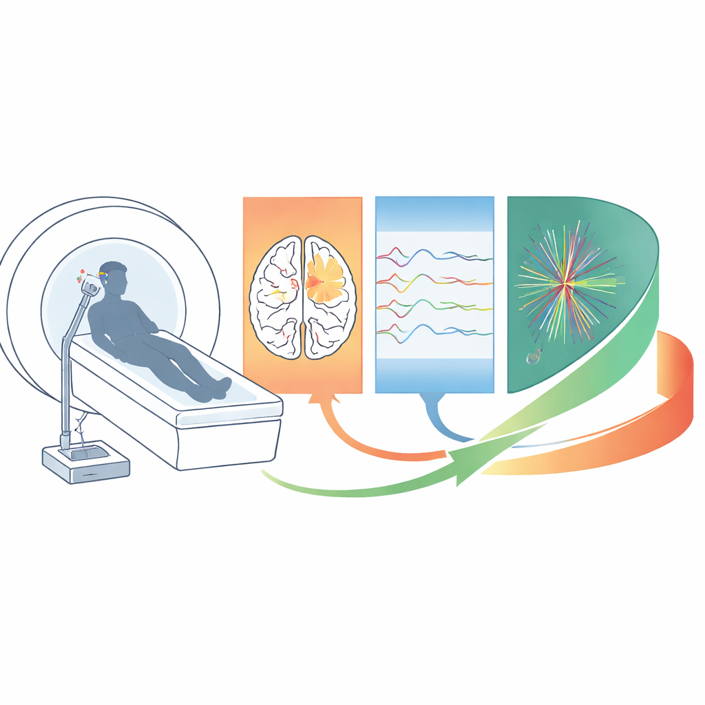
No single brain scanner can see everything that happens when attention wavers. Functional MRI (fMRI) shows where activity changes across the brain in fine spatial detail, but it is relatively slow. Electroencephalography (EEG) tracks rapid electrical activity on the scale of milliseconds, but blurs where in the brain the signals come from. In this study, 28 healthy adults lay in an MRI scanner wearing an EEG cap so that both kinds of data were collected at the same time. The team also added diffusion imaging, which maps the brain’s wiring, allowing later studies to relate fast electrical changes, slower blood-flow changes, and the underlying white‑matter connections that link brain regions.
A daydream-inducing attention challenge
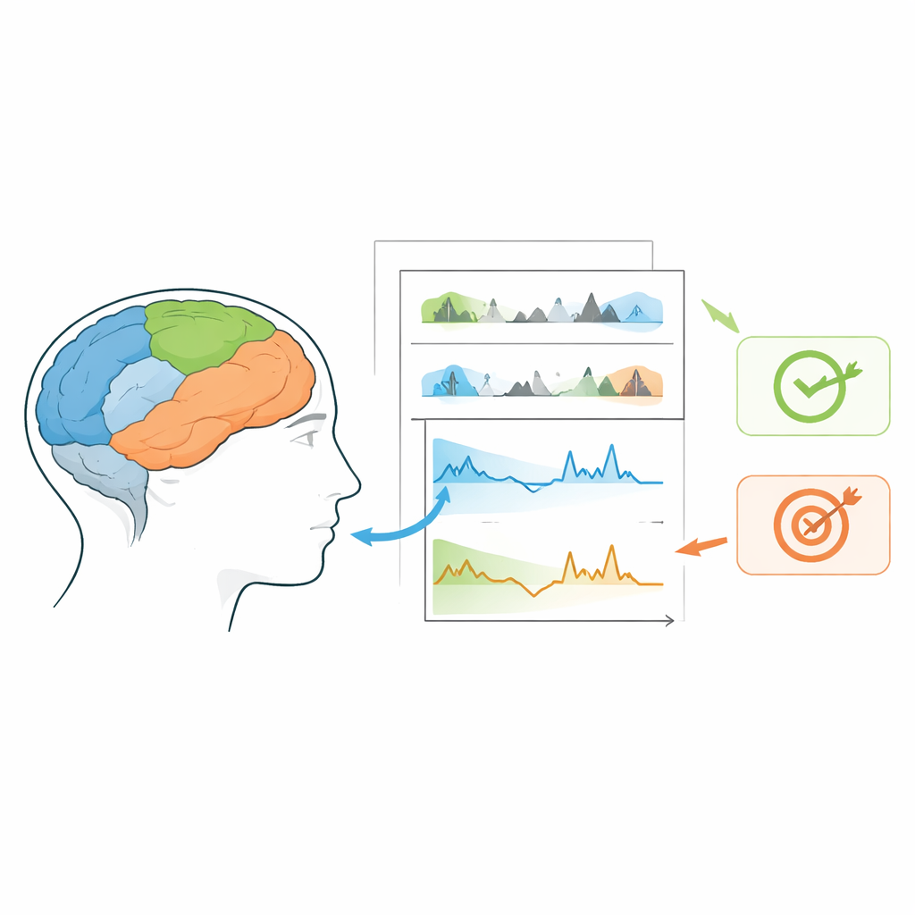
The central experiment uses a gradual continuous performance task called gradCPT, which is especially good at revealing moment-to-moment changes in focus. Participants view a stream of city and mountain scenes that gently fade into one another every 800 milliseconds. They must press a button for most city images but hold back for the less frequent mountain scenes. This simple rule makes lapses obvious: a missed button press or a mistaken press on a mountain marks a slip in attention. To probe how inner thoughts interfere with the task, the researchers also included blocks where people were asked to vividly imagine their future work or school day while still doing gradCPT, as well as periods of pure imagination, a flickering checkerboard visual task, and resting with eyes open or closed.
From noisy signals to usable brain maps
Recording EEG inside an MRI scanner is technically challenging: the powerful magnetic fields and rapid switching gradients create large electrical artifacts that can overwhelm the tiny brain signals of interest. The team carefully prepared each participant, minimized cable movement, and followed best‑practice layouts for amplifiers and wiring. They then used specialized software to subtract repeating MRI-related noise patterns, remove heartbeat-related distortions, and filter out other artifacts. For the fMRI data, they applied a modern, standardized preprocessing pipeline, corrected for head motion and scanner distortions, smoothed the images, and quantified signal quality. Diffusion images were cleaned and used to reconstruct major white‑matter pathways and connectivity matrices between over a hundred brain regions. Quality checks showed that motion was low, key signals were robust, and structural connections looked anatomically plausible.
How brain networks hint at upcoming errors
To demonstrate what can be done with the dataset, the authors repeated a classic finding about attention lapses. They focused on three major brain networks: visual regions that process the scenes, attention networks that support goal‑directed focus, and the “default mode” network, which becomes more active during inward‑focused thought and mind‑wandering. When they compared brain activity before correct versus incorrect trials in the gradCPT, they found that errors were preceded by stronger activity in default‑mode areas and weaker activity in visual areas. This pattern was especially clear during “out‑of‑the‑zone” periods, when reaction times fluctuated more and behavior was less stable. In other words, the brain was already shifting toward internal thoughts and away from the visual task moments before performance slipped.
A shared resource for studying focus and distraction
Beyond these initial demonstrations, the real product of this work is the public dataset itself. Organized in a widely used format and shared on the OpenNeuro platform, it includes raw and preprocessed EEG, fMRI, diffusion imaging, and behavioral data, plus code for task presentation and analysis. Researchers can use it to test new methods for cleaning EEG inside MRI, explore how structural wiring supports attention, or build models that predict lapses from patterns across multiple brain signals. For non‑specialists, the take‑home message is straightforward: when our minds wander, the balance between brain networks that look outward and those that turn inward shifts in detectable ways—and this dataset offers a powerful new window into how those shifts unfold over time.
Citation: Cha, Y., Lee, Y., Ji, E. et al. Sustained Attention Task (gradCPT) Dataset using simultaneous EEG-fMRI and DTI. Sci Data 13, 573 (2026). https://doi.org/10.1038/s41597-026-06616-6
Keywords: sustained attention, mind wandering, EEG fMRI, brain networks, open neuroimaging data