Clear Sky Science · en
Functional bipartite invariance in mouse primary visual cortex receptive fields
How the Brain Sees Through Visual Clutter
Finding a friend in a crowd or spotting a bird in leafy branches feels effortless, but it demands a remarkable skill: our brains must recognize important things even as lighting, distance, and background constantly change. This paper explores how tiny circuits in the mouse brain achieve such flexible vision. The authors uncover an unexpected way that individual neurons split their “view” of the world into two parts to stay sensitive to object boundaries while ignoring distracting details.
Probing the Visual Brain with Smart Computers
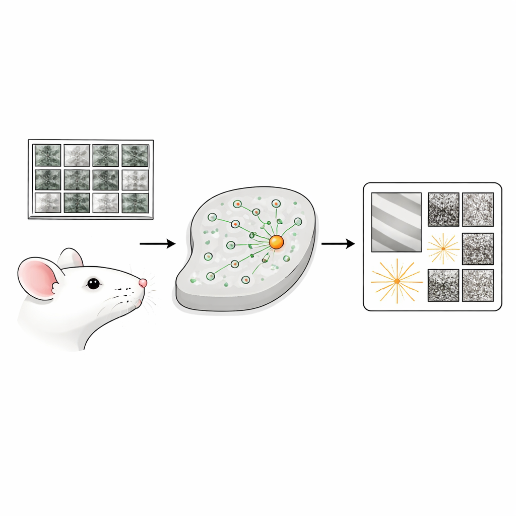
The study focuses on the primary visual cortex, a region that first processes images from the eyes. Instead of presenting simple patterns like stripes, the researchers showed awake mice thousands of rich, natural photographs while recording the activity of tens of thousands of neurons. They then trained a deep-learning model to predict how each neuron would respond to any new image. This model served as a “digital twin” of the real brain tissue: a fast, flexible stand-in that could be probed with countless synthetic images that would be impractical to test directly in animals.
Many Different Pictures, Same Neuron Excitement
Using the digital twin, the team generated, for each neuron, a most exciting image (its favorite picture) and then a set of “varied exciting inputs.” These are 20 images that look as different as possible from one another yet all strongly activate the same cell. When they showed these special images back to the mice, the real neurons lit up almost as strongly as the model predicted, confirming that the synthesis method had uncovered genuine properties of the brain. The patterns across these images summarized what each neuron cared about and which changes in the image it could tolerate without losing its enthusiasm.
A Surprising Split View Inside Single Neurons
Many neurons behaved in expected ways, responding to simple edges or to textures that could shift around without changing the response. But a large group displayed a new kind of behavior the authors call “bipartite invariance.” For these neurons, the patch of visual space they monitor effectively splits into two nonoverlapping zones. In one zone, the neuron demands a specific, fixed pattern. In the other zone, it responds robustly as long as some version of a preferred fine-grained texture appears there, even if that texture is shifted around. This combination—a rigid pattern next to a flexible texture detector—cannot be explained by classic models of early visual cells and suggests that these neurons are specially tuned to transitions between different kinds of surface detail.
Detecting Object Edges in a Noisy World
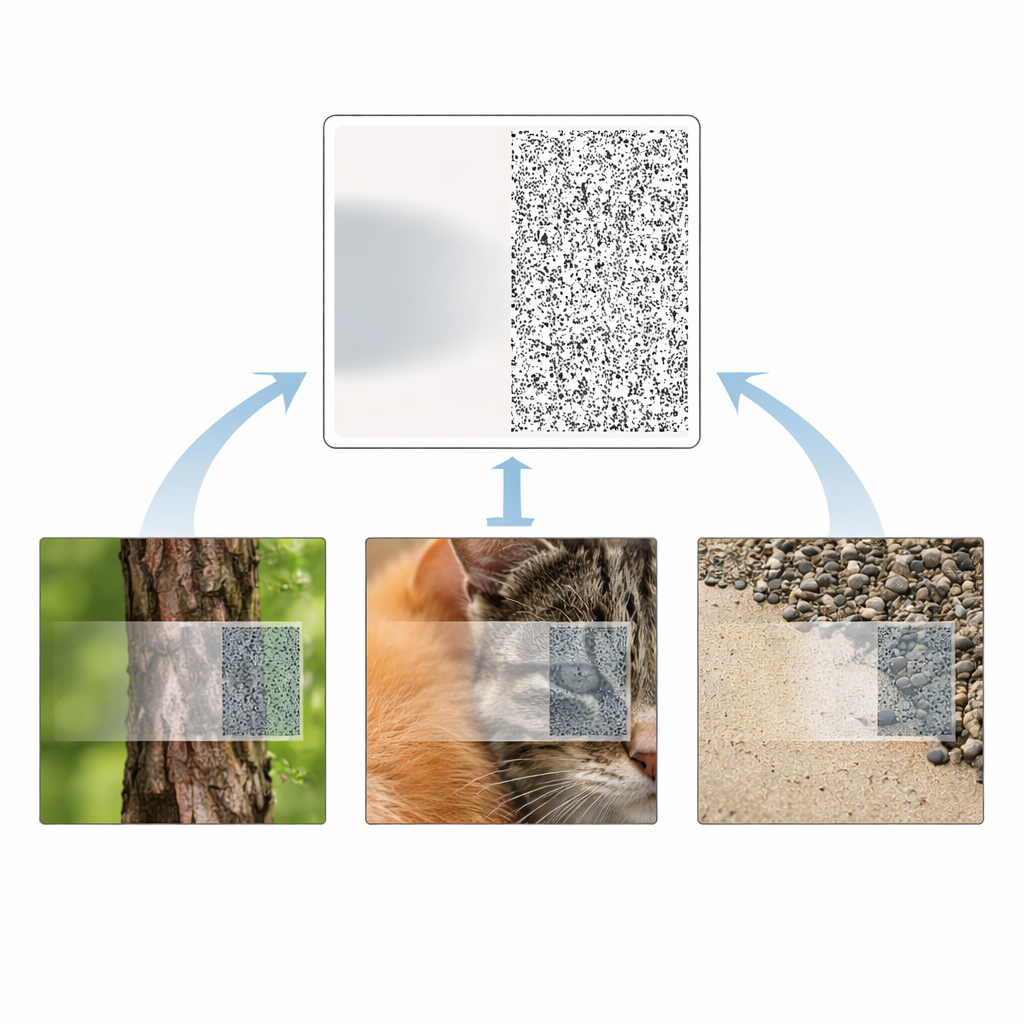
The researchers next asked what this split structure might be good for in real scenes. They scanned more than a million labeled bird photographs, looking for natural crops that strongly activated each neuron. Those highly effective crops tended to contain the edge between bird and background. Moreover, the side of the neuron’s receptive field that tolerated variable input typically favored high spatial detail—fine feathers, bark, or gravel—while the fixed side preferred smoother, lower-frequency patterns. When the team rebuilt the bird images using simple gratings, they found that these neurons strongly preferred boundaries where the two sides differed in texture sharpness, sometimes along with orientation differences. In other words, these cells seem wired to flag places where the visual world changes abruptly from coarse to fine structure, a powerful cue for separating objects from their surroundings.
Connecting Circuit Wiring to Flexible Vision
To relate this computation to physical wiring, the authors turned to a massive “functional connectomics” dataset in which both the responses and synaptic connections of thousands of mouse visual neurons have been mapped. Applying their digital-twin approach there, they found that neurons receiving input (postsynaptic cells) generally showed more invariance—greater tolerance to varied exciting inputs—than the neurons feeding into them. At the same time, neurons with lower invariance tended to form more connections. This pattern supports a long-standing idea: more complex, tolerant responses are built by pooling signals from many simpler inputs, but here this hierarchy is demonstrated within a single cortical layer, not just between brain areas.
Why This Matters for Brains and Machines
Together, these findings reveal a new organizing principle in the mouse visual cortex: many neurons carve their view into one stable region and one texture-flexible region, making them natural detectors of boundaries shaped by differences in fine detail. This bipartite structure appears robust across methods and aligns neatly with real-world object edges, offering a concrete circuit-level mechanism for how the brain segments cluttered scenes. Beyond basic neuroscience, the work also suggests design ideas for artificial vision systems, where introducing similar split-field units could make computer vision more robust to background changes while keeping sharp sensitivity to object outlines.
Citation: Ding, Z., Tran, D., Ponder, K. et al. Functional bipartite invariance in mouse primary visual cortex receptive fields. Nat Neurosci 29, 851–863 (2026). https://doi.org/10.1038/s41593-026-02213-3
Keywords: visual cortex, texture segmentation, neuronal invariance, object boundaries, computational neuroscience