Clear Sky Science · en
PinkyCaMP: an mScarlet-based calcium sensor with enhanced brightness, photostability and multiplexing capabilities
Seeing the Brain’s Hidden Sparks
Every thought, memory and movement in your body depends on tiny flashes of calcium inside brain cells. Scientists track these calcium bursts using glowing molecules, turning invisible nerve activity into movies of light. But the red versions of these glowing sensors have long been dim, fragile and tricky to use alongside other light-based tools. This study introduces PinkyCaMP, a new bright pink sensor designed to make watching the brain’s activity clearer, safer and easier to combine with modern optical methods.
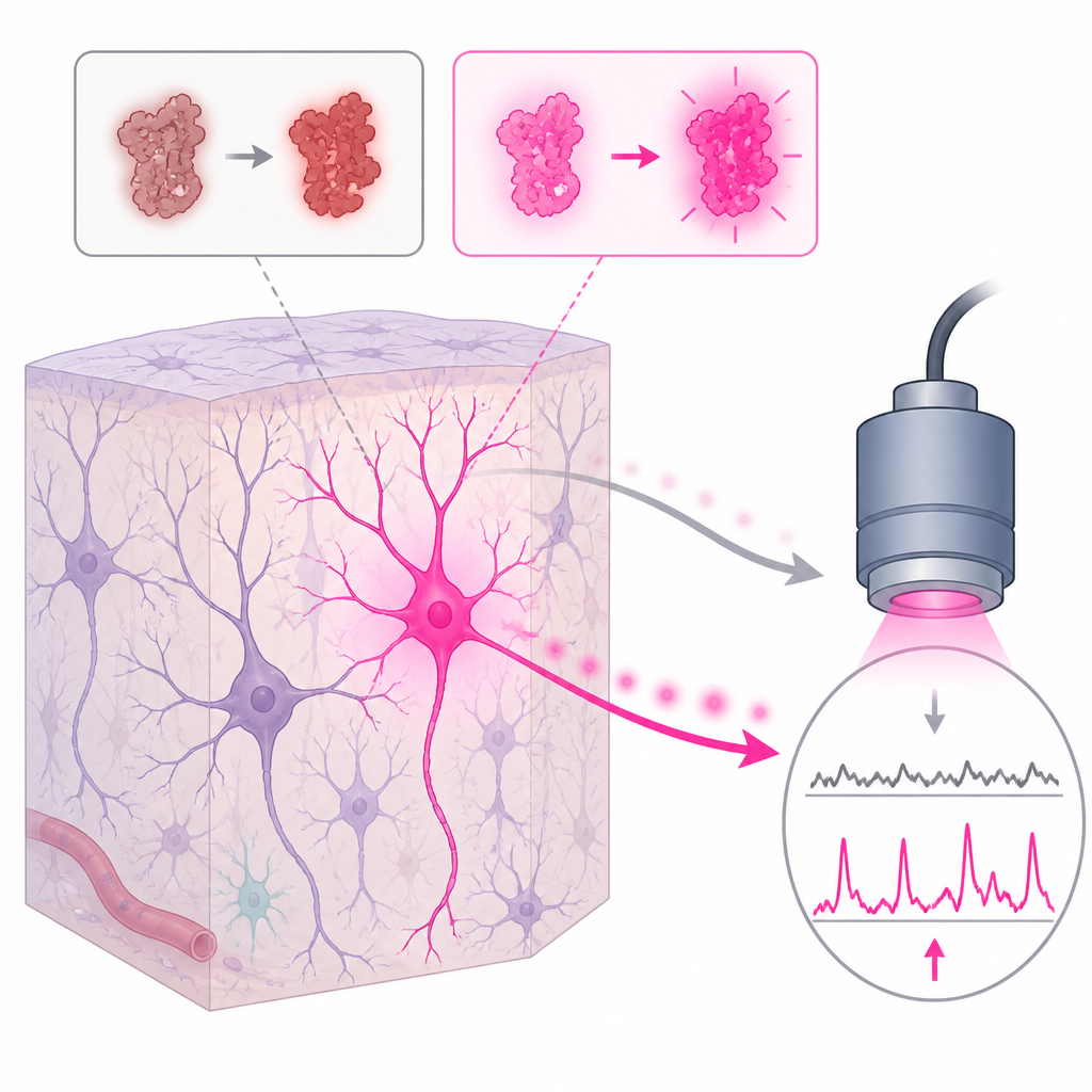
Why Brighter Brain Movies Matter
Neuroscientists often label neurons with special proteins that light up when calcium levels rise, signaling that a cell is active. Green sensors already work very well, but red ones have important advantages: red light penetrates deeper into tissue, causes less damage and can be separated more easily from blue light used to control neurons with optogenetics. Existing red sensors, however, tend to be too dim, fade quickly under the microscope and can respond falsely when exposed to blue light. These weaknesses limit experiments that need to both control and record from brain cells, or to follow several signals at once in the same animal.
Building a Better Pink Sensor
To tackle these problems, the researchers built a new sensor using mScarlet, one of the brightest known red fluorescent proteins. They re-engineered its structure so that the protein’s glow would change whenever it binds calcium. This involved cutting and reconnecting parts of the molecule, and surrounding it with calcium-sensing components drawn from earlier sensors. The team then created thousands of variants and screened them for both brightness and responsiveness. After twelve rounds of fine-tuning, they selected a version that balanced intense glow with strong calcium sensitivity and stability, and named it PinkyCaMP.
Measurements on purified protein showed that PinkyCaMP shines much more brightly than previous red sensors when calcium is present, while remaining relatively calm when calcium is low. It also withstands long periods of illumination without bleaching as quickly. Importantly, tests confirmed that Blue light, often used to drive optogenetic switches, does not trigger false signals in PinkyCaMP, solving a major source of confusion in earlier red tools.
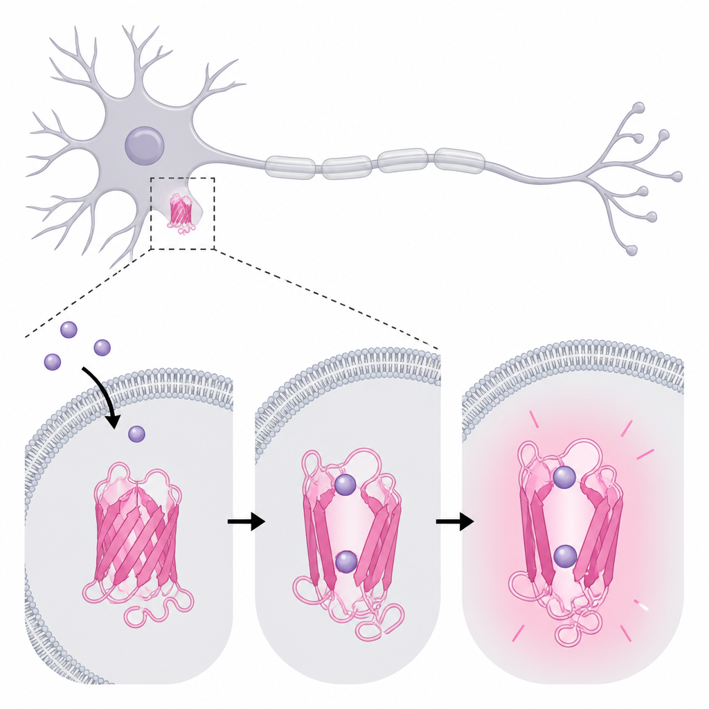
Putting PinkyCaMP to Work in Brain Cells
The researchers next tested PinkyCaMP in living cells and brain tissue. In cultured human cells, PinkyCaMP glowed several times brighter than leading red sensors. In mouse neurons grown in dishes, the pink signal closely tracked electrical spikes, and the size of the light flashes grew reliably with stronger stimulation. Compared to older red indicators and a popular green one, PinkyCaMP produced the strongest overall signals and kept working under continuous light with less fading. It also avoided clumping inside cellular waste compartments, a chronic issue that weakens many red sensors.
In brain slices from mice, PinkyCaMP recorded spontaneous bursts of coordinated activity with high signal-to-noise ratio, meaning real events stood out clearly from background fluctuations. When directly compared with a recent bright red sensor, PinkyCaMP worked well even under gentler light, and still outperformed its rival under harsher conditions, giving larger and cleaner responses before fading. These tests suggest that researchers can use lower light levels, reducing the risk of damaging tissue while still getting usable data.
Watching Behavior and Chemistry in Living Mice
To see how PinkyCaMP performs in complex, moving animals, the team expressed it in specific brain regions of mice. Using thin optical fibers, they monitored calcium signals in the prefrontal cortex while mice experienced a brief puff of air or explored a maze that tests anxiety. PinkyCaMP reported strong, reliable activity surges that matched the animals’ behavior, whereas control fluorescent proteins did not change. By pairing PinkyCaMP with a separate green sensor for serotonin, they could simultaneously track how brain cell firing and a key mood-related chemical responded to the same stressful event, all through one implanted fiber.
The sensor also proved compatible with blue-light optogenetics. In one experiment, PinkyCaMP recorded how brain cells in a memory-related region became more active when nearby inhibitory cells were switched off with a blue-light-sensitive channel. Control animals that lacked this switch showed no such change, confirming that the pink signals reflected real circuit activity rather than an artifact of illumination. In addition, PinkyCaMP worked well with advanced imaging setups, including conventional two-photon microscopes and tiny head-mounted devices, allowing recordings from awake, head-fixed or freely moving mice over weeks to months.
What This Means for Future Brain Research
Taken together, the results show that PinkyCaMP narrows the gap between red and green calcium sensors. It offers brightness, durability and cleaner signals than previous red tools, while avoiding misleading responses to blue light. Although its relatively slow relaxation makes it less suited to tracking extremely rapid firing patterns, this same high sensitivity makes it ideal for following sparse or subtle activity across many cells and deep brain regions. Because PinkyCaMP can be used alongside green indicators and blue-light optogenetics, it opens the door to richer, multicolored views of how different cell types and chemical signals work together in the living brain.
Citation: Fink, R., Imai, S., Gockel, N. et al. PinkyCaMP: an mScarlet-based calcium sensor with enhanced brightness, photostability and multiplexing capabilities. Nat Methods 23, 998–1010 (2026). https://doi.org/10.1038/s41592-026-03065-2
Keywords: calcium imaging, fluorescent sensor, optogenetics, neuronal activity, two photon microscopy