Clear Sky Science · en
Vectorized instructive signals in cortical dendrites
How Brains Learn from Their Own Neurons
Modern artificial intelligence programs learn by carefully adjusting millions of tiny connections so that each neuron in the network gets its own teaching signal. Neuroscientists have long wondered whether the biological brain has a comparable way to tell individual neurons how to change during learning. This study shows that the answer is yes: mice appear to use special signals in the thin branches of their brain cells to guide learning, offering a biological counterpart to core ideas in machine learning.
Turning Thought into a Moving Picture
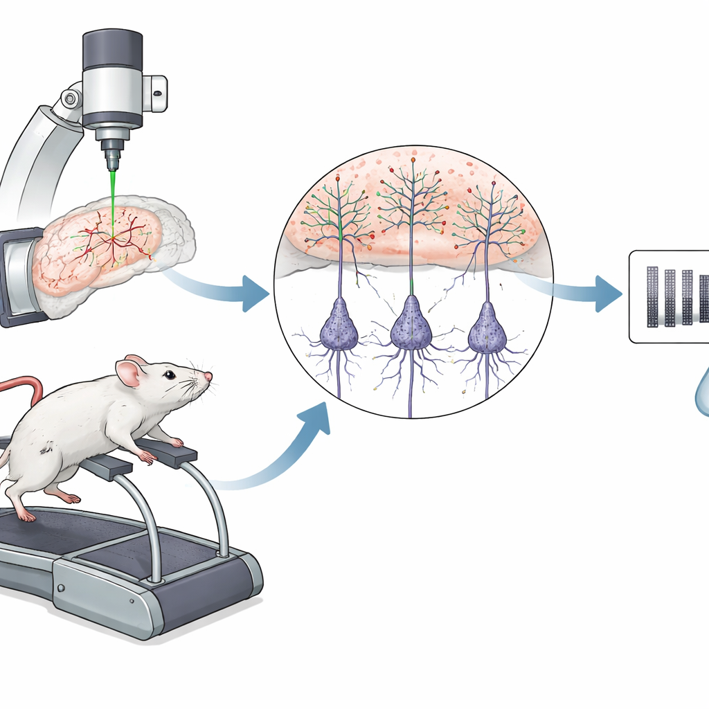
To make the learning process visible, the researchers built a brain–computer interface for mice. While a mouse ran on a small treadmill, a microscope peered into a part of its brain involved in navigation and memory. The scientists chose two tiny sets of neurons in this region and linked their activity to a drifting striped pattern on a screen. When one group of neurons became more active than the other, the pattern rotated toward a specific “target” orientation, and the mouse received a sweet reward. In this way, the experimenters defined a precise rule that tied each neuron’s activity to success or failure.
Two Populations, Opposite Jobs
The two neuron groups were intermingled in the same patch of cortex, but they had opposite effects on the stimulus: one pushed the pattern toward the rewarding angle, the other pushed it away. Over about two weeks of training, mice learned to earn more rewards per minute and to succeed on a higher fraction of trials. At the same time, the “helpful” neurons tended to keep firing strongly, whereas the “unhelpful” ones gradually fired less. This shift did not simply reflect a global change in arousal or running speed; instead, it matched how each neuron’s activity influenced task performance, hinting that the brain was solving a fine-grained credit assignment problem.
Listening to the Hidden Ends of Neurons
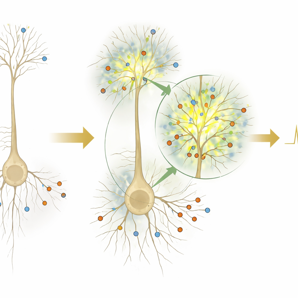
Crucially, the team looked not only at the main bodies of these neurons but also at their long, tree-like branches called apical dendrites. These branches sit closer to the brain’s surface and receive different inputs than the cell body does. By rapidly switching focus between deep and superficial imaging planes, the researchers recorded activity from both sites in the same neuron. They found that when a neuron fired, the strength of the signal in its dendrites could be either larger or smaller than expected from the signal at the soma, and that these mismatches were partly predictable from the activity pattern of nearby neurons. This means the dendrites were not just passively echoing the soma; they were receiving distinct information from the surrounding network.
Dendrites Carry Reward and Error Signals
The next question was what those dendritic signals meant. By analysing the timing of events relative to reward delivery and trial outcome, the authors showed that patterns of dendritic amplification across many neurons could be used to tell rewarded from unrewarded trials, and successful from unsuccessful attempts, even before the outcome occurred. In other words, the dendritic signals carried information about both reward and ongoing task error that could, in principle, serve as teaching signals. Strikingly, the sign of these signals depended on the neuron’s causal role: dendrites of neurons that helped reduce error were boosted when error decreased, while dendrites of neurons that pushed the pattern in the wrong direction were boosted when error increased. This “vectorized” pattern mirrors how artificial networks send different error messages to different units.
Shutting Down the Teaching Channel
To test whether these dendritic patterns mattered for learning, the researchers used light-sensitive proteins to activate a specific class of inhibitory cells that sit in the outermost layer of cortex and target apical dendrites. Turning on these inhibitory cells strongly weakened the dendritic mismatches relative to somatic activity. Under these conditions, the population of dendrites no longer reliably signaled reward or trial outcome, and mice failed to improve at the brain–computer task, even though the basic setup was unchanged. Similar effects occurred under anesthesia, when top-down inputs are known to be dampened, further supporting the idea that these dendritic signals depend on specialized feedback pathways.
What This Means for Understanding Learning
Taken together, the findings suggest that the brain uses separate electrical compartments within single neurons to route teaching signals, much like how artificial networks direct error signals to particular units. The apical dendrites of cortical neurons appear to carry neuron-specific information about reward and error changes, and interfering with this channel blocks learning. For a non-specialist, the key message is that learning in the brain may hinge not just on which neurons are active, but on how their hidden branches integrate feedback from the rest of the network—providing a biological blueprint for some of the core ideas behind modern machine learning.
Citation: Francioni, V., Tang, V.D., Toloza, E.H.S. et al. Vectorized instructive signals in cortical dendrites. Nature 652, 1254–1263 (2026). https://doi.org/10.1038/s41586-026-10190-7
Keywords: dendritic computation, credit assignment, brain–computer interface, cortical learning, neurofeedback