Clear Sky Science · en
Ultrafast holographic chiroptical microscopy
Seeing Hidden Twists in Materials
Many of today’s most promising technologies, from better solar cells to faster computers, rely on how tiny particles inside materials twist, spin, and move in billionths of a second. Until now, scientists could either measure these ultrafast twists in a single averaged spot or take static pictures over a wide area, but not both at once. This paper introduces a new kind of microscope that can film these fleeting changes in polarization – the way light’s electric field points – across a whole miniature landscape, opening a window into hidden patterns of magnetism and electronic motion.
A New Way to Watch Light Interact with Matter
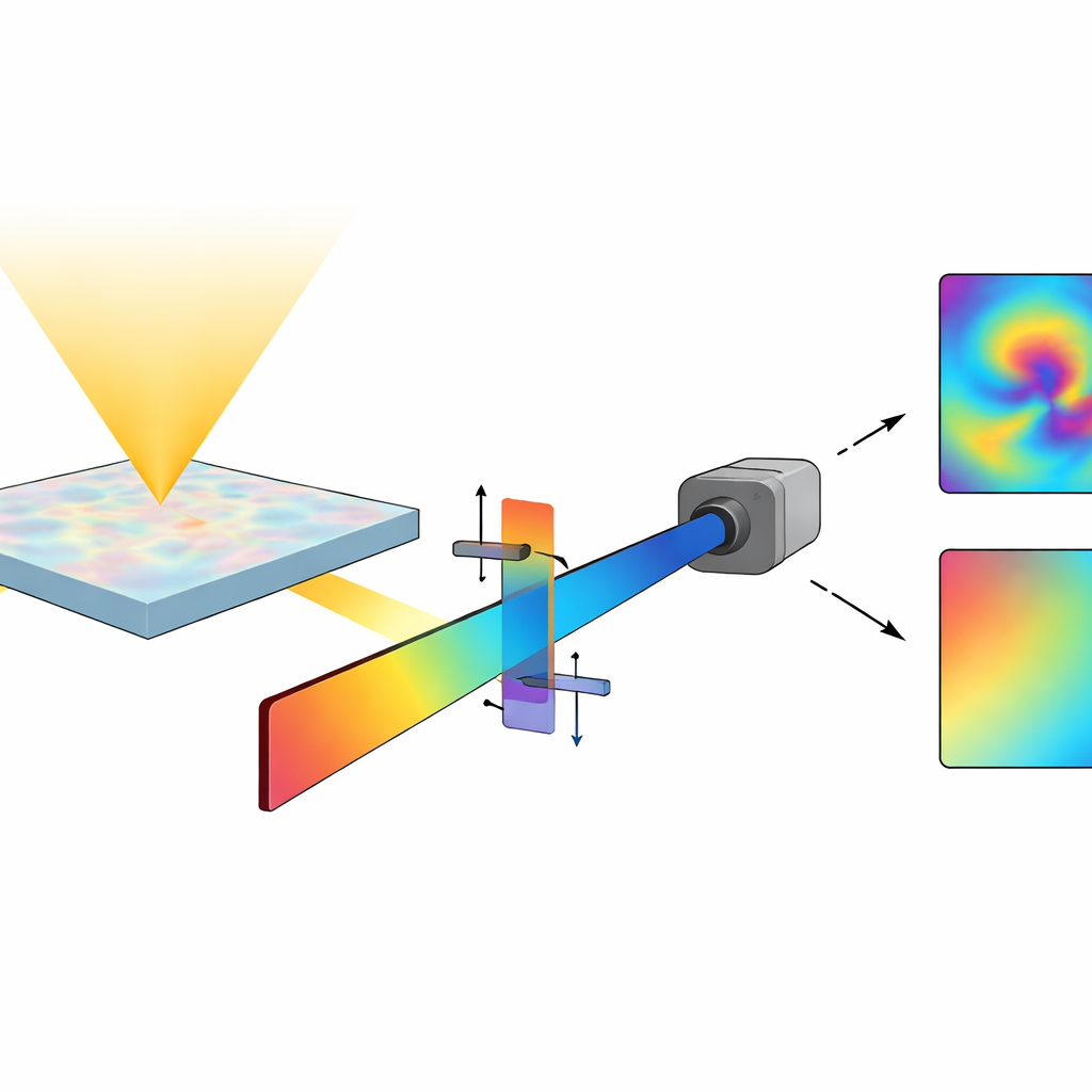
The researchers set out to solve a long-standing problem: how to image “chiral” responses – tiny differences in how a material reacts to left- versus right-handed light – over a large field of view and on femtosecond timescales (a femtosecond is a millionth of a billionth of a second). Traditional methods could detect these effects very sensitively, but only by averaging over a big area, wiping out local details. The new instrument combines a widefield microscope with a holographic trick that lets the camera capture not just how bright the light is at each pixel, but also how its polarization has rotated or become more elliptical as it passes through the sample.
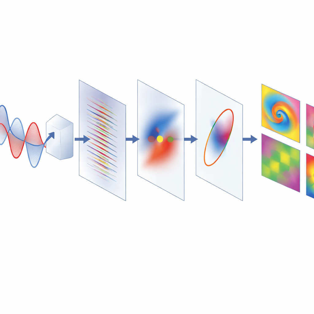
How Holograms Capture Polarization Movies
At the heart of the setup is a “pump–probe” experiment. A first pulse of light (the pump) briefly disturbs the material, changing the spins and charges inside it. A second pulse (the probe) then passes through the sample and carries away information about how it has been altered. Instead of recording this probe pulse directly, the microscope makes it interfere with two carefully arranged reference pulses whose polarizations are at right angles to each other. Because these references arrive on the camera at a slight angle, they create interference patterns with distinct stripe directions for the horizontal and vertical components of the probe. By taking a spatial Fourier transform of the recorded hologram and picking out the right stripes, the team reconstructs, at every pixel, the full electric field of the probe in two directions, including its phase. From this, they can compute maps of how much light is absorbed, how much its phase is shifted, and how its polarization ellipse twists and stretches.
Mapping Spins and Bandgaps in Perovskite Films
To showcase the power of the technique, the authors study hybrid perovskites, a family of semiconductors central to next-generation solar cells and light emitters. In these materials, strong coupling between electrons and their spins allows circularly polarized light to create spin-polarized excitons, whose presence slightly rotates the polarization of a passing probe beam. With the new microscope, they directly visualize how this rotation and related signals change in space and time. In a bromide-only perovskite, they see micrometer-scale regions where the probe’s transmission for the two orthogonal polarizations changes with opposite signs, as expected from a transient rotation that decays over just a few trillionths of a second, revealing the spin lifetime. Phase images, sensitive to how the refractive index changes, track slower processes such as the cooling of “hot” carriers and their long-lived population.
Revealing Hidden Domains and Spin Transport
In a mixed-halide perovskite, the microscope uncovers a patchwork of domains where the rotation signal alternates between positive and negative values, while the phase signal also varies but does not flip under reversal of the pump’s circular polarization. This pattern points to local variations of the electronic bandgap caused by subtle changes in composition, not to true magnetic domains – information that would be lost in a conventional bulk measurement. In a second experiment, the team structures the pump light into an array of diffraction-limited spots and uses the transient rotation signal to watch how spin-polarized carriers spread out from each spot. By following how the width of the rotation pattern grows over time at different excitation strengths, they extract diffusion behavior and how it speeds up when carriers scatter more strongly at higher densities.
Why This Matters for Future Materials
For a non-specialist, the key message is that this work turns what used to be a single number – an average chiral signal – into detailed movies that show where and how spins and charges move in complex materials. The microscope combines sensitivity close to fundamental noise limits with sharp spatial and temporal resolution, and it delivers both chiral and non-chiral information from the same dataset. Because the approach is general, it can now be applied to systems ranging from biomolecules and chiral nanostructures to emerging spintronic and topological materials, helping researchers design devices that exploit the twist and spin of light and matter in ever more precise ways.
Citation: Hörmann, M., Visentin, F., Gessner, J.A. et al. Ultrafast holographic chiroptical microscopy. Nat. Photon. 20, 592–599 (2026). https://doi.org/10.1038/s41566-025-01824-9
Keywords: ultrafast microscopy, chiroptical imaging, perovskite semiconductors, spin dynamics, holographic imaging