Clear Sky Science · en
Glycan atlassing enables functional tracing of cell state
A sugar coat that tells cell stories
Every cell in your body wears a sugar-rich coat that quietly shapes how it behaves and how it talks to its neighbors. This study shows that by zooming in on that coat with extraordinary detail, scientists can read out whether a cell is healthy, turning cancerous, firing as a neuron or reacting as an immune cell. The work introduces a way to turn this delicate sugar layer into a practical readout of cell state, with implications for cancer diagnosis, brain research and immune therapies.
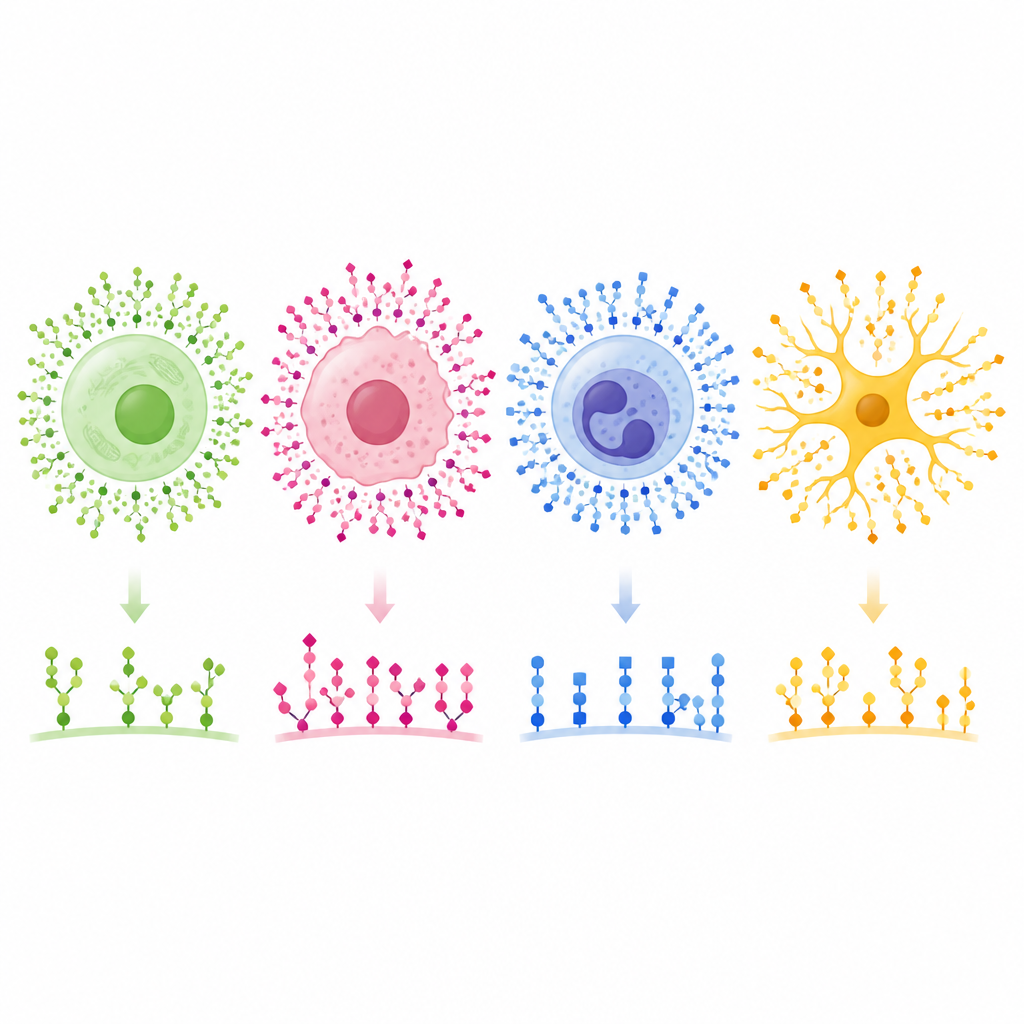
Seeing the hidden sugar shell
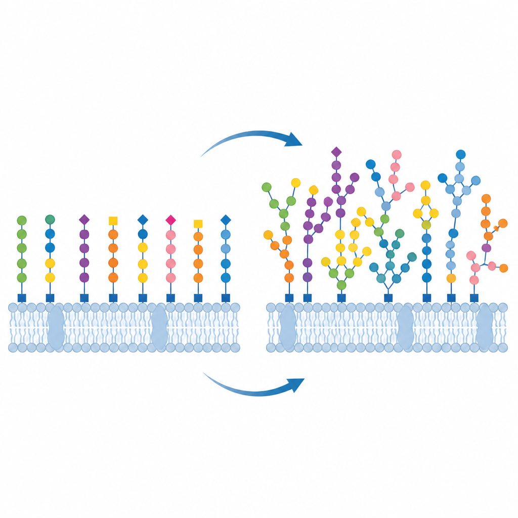
The outer coat of cells, called the glycocalyx, is made of countless sugar chains attached to fats and proteins in the membrane. These chains come in many shapes and sizes, and their exact arrangement is thought to influence processes such as immune recognition, tissue growth and how pathogens gain entry. Traditional tools can identify which sugars are present, but not how they are arranged in place on intact cells. Electron microscopes give sharp images but can disturb this fragile layer, and standard light microscopes cannot resolve features smaller than about one quarter of a micrometre. What has been missing is a way to map the fine structure of this sugar coat on real cells while also tying those patterns to what the cell is actually doing.
Turning sugars into a nanometre atlas
The authors developed a technique they call glycan atlassing, which converts the surface sugars into a high resolution map. They first label different sugar motifs using lectins, natural proteins that stick to specific sugar shapes, each carrying a short DNA tag. They also feed cells specially designed sugar building blocks that get incorporated into certain surface sugars and then linked to DNA tags using a gentle click chemistry reaction. During imaging, short fluorescent DNA strands temporarily bind and unbind to these tags, creating single molecule blinks that can be localized with nanometre precision. By cycling through multiple DNA codes, they capture several sugar types in the same cell without blurring them together.
Finding patterns in dense sugar forests
Collecting such ultra sharp images is only half the story; the other half is making sense of the dense constellations of points. The team built an analysis pipeline that first groups repeated blinks into single “binding sites” for each lectin, then measures how close each site is to its nearest neighbors across all sugar types. They also use a software tool called GlyCo to cluster sites that lie within a few nanometres of each other, which likely belong to the same sugar chain or tiny cluster. From these distances and groupings, they extract characteristic spatial signatures and feed them into a statistical method called principal component analysis that can separate different cellular conditions based on their sugar organization alone.
Reading cancer, brain and immune states
To show what glycan atlassing can do, the researchers applied it to a range of systems of rising complexity. In a breast cell model, it distinguished normal cells, oncogene driven cells and cells pushed through an early step of cancer spread called epithelial to mesenchymal transition. The sugar coat changed not through a single dramatic shift, but through many subtle rearrangements that together marked each stage. In developing rat neurons, the method picked up differences between cell bodies and branching extensions that matched known timing of sugar maturation, hinting at a link between local sugar patterns and neuron function. In human immune cells, including natural killer cells, CD4 T cells and neutrophils, the sugar coat remodelled within minutes after activation, revealing a fast and previously underappreciated layer of immune regulation. Finally, in slices of human breast tumors, glycan atlassing separated tumor regions from nearby non tumor tissue based solely on their nanoscale sugar signatures, with cancerous zones showing more varied and disordered patterns.
From sugar fingerprints to future medicine
Overall, the study shows that the fine scale layout of the cell’s sugar coat carries rich information about what state the cell is in, from early cancer changes to immune activation and tissue health. Glycan atlassing turns this outer layer into a measurable fingerprint, raising the possibility of classifying tumors, tracking immune therapies or probing brain function by reading sugar patterns rather than bulk markers alone. While the method still requires specialized labels and expertise, it points toward a future where doctors and researchers may routinely use nanoscale sugar maps to understand disease and guide treatment.
Citation: Moonnukandathil Joseph, D., Yurekli, N., Fritsche, S. et al. Glycan atlassing enables functional tracing of cell state. Nat. Nanotechnol. 21, 720–731 (2026). https://doi.org/10.1038/s41565-026-02151-y
Keywords: glycocalyx, cell surface sugars, super resolution microscopy, cancer glycosylation, immune cell activation
See more on the researcher's website: https://mpl.mpg.de/research-at-mpl/independent-research-groups/moeckl