Clear Sky Science · en
Spatial transcriptomics maps host–gut microbiome biogeography at high resolution
Peering Into the Hidden City in Our Guts
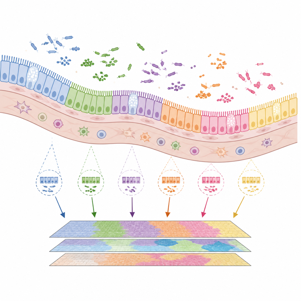
Inside every one of us lives a bustling city of microbes that help digest food, train the immune system and may even influence cancer. Yet most tools for studying this inner world blur away fine details, like looking at a city only from space. This study introduces a way to map where different bacteria sit in the gut, how they cluster together and how they line up against our own cells, all at nearly single-microbe resolution. The work offers a sharper view of how the microbiome is built and how it changes near tumours in the intestine.
A New Way to See Microbes in Place
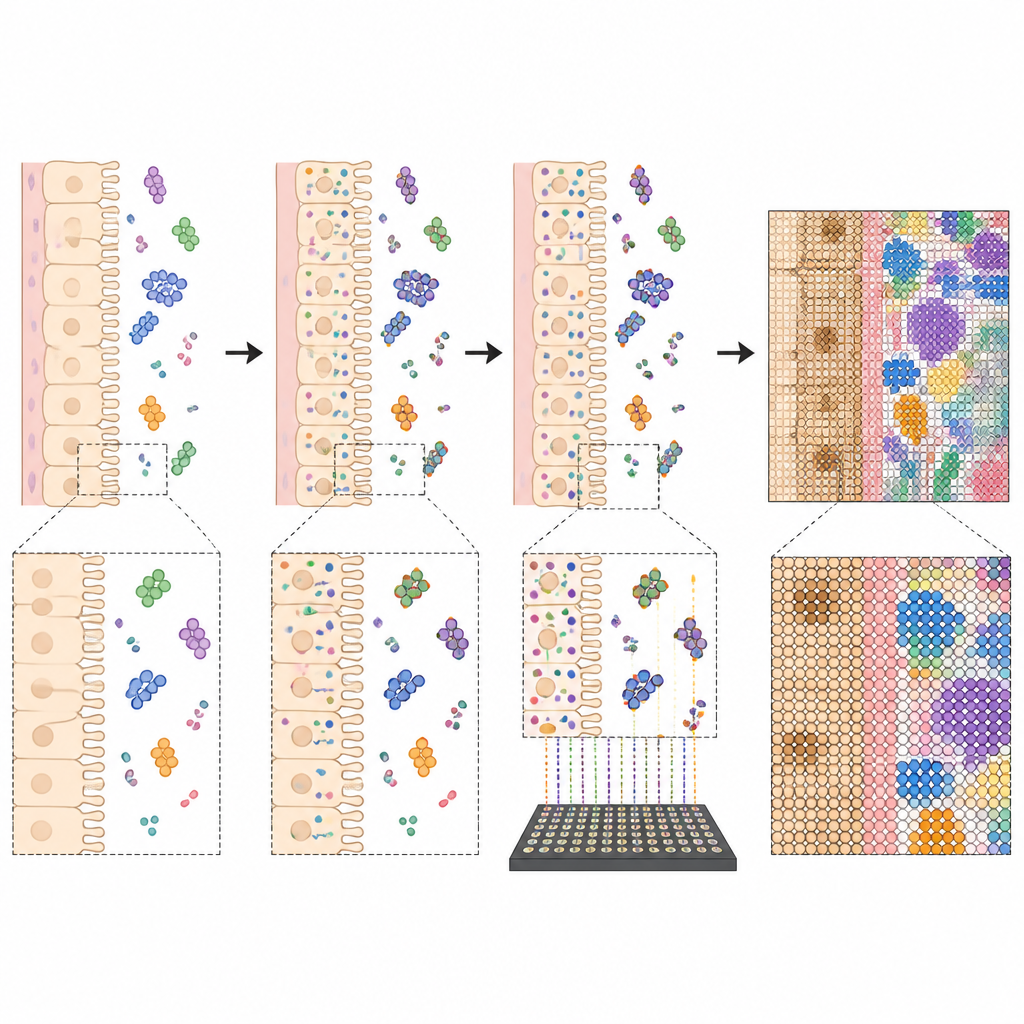
The authors build on a technology called spatial RNA sequencing, which reads out which genes are active in thin slices of tissue while keeping track of where each signal came from. Standard versions of this method work well for our own cells but largely ignore bacteria, whose RNA lacks a particular tail that these tools are designed to catch. The team solved this by adding an extra enzyme step that glues tails onto both microbial and host RNA directly inside the tissue slice. This simple tweak allowed common commercial platforms to pick up microbial messages up to about one hundred times more efficiently without losing information from host genes.
Tracing Microbial Neighborhoods Along the Gut
Using mice, including a model of intestinal cancer, the researchers applied their method to several regions of the gut, from the upper small intestine to the colon. At lower magnification they mapped how many kinds of bacteria appeared in each tiny spot and how this changed from the tissue wall toward the open lumen. As expected, the large intestine hosted a richer mix of bacterial families than the small intestine. Some groups were found mostly in the centre of the gut contents, while others preferred to live nearer the mucosal surface where they sit closer to host cells. These broad patterns matched previous bulk measurements but now came with fine-grained spatial detail.
Zooming in to Single-Cell Scales
The team then used a higher resolution platform to reach pixel sizes of about half a micrometre, small enough to resolve individual bacteria and the architecture of the gut lining. They could assign host RNA to single cells, identify cell types, and overlay this with the positions of microbial RNA. The maps revealed that mature absorptive cells near the tips of the villi were highly active, while stem-like cells in the crypts showed more newly made, unspliced RNA. On the microbial side, many genera formed distinct colonies whose sizes varied from just a few micrometres to over thirty. By analysing how these clusters were arranged, the authors inferred frequent short-range interactions within and between bacterial groups, painting a picture of a highly structured microbial ecosystem rather than a uniform soup.
Microbes at the Edge of Tumours
Because cancers in the gut arise at barrier surfaces, the team asked how the local microbiome reorganizes around tumours. In healthy regions, bacteria tended to be densest a short distance away from the villi, separated from the tissue by mucus and other defences. Near tumours, however, the peak density of bacteria shifted right up against the tumour boundary. Key genera that normally sit further from the wall now hugged the cancer edge, and tumour zones were enriched in rapidly dividing intestinal cells and immune cells. These findings suggest that both the reshaped tissue structure and the changing cell mix around a tumour can pull microbial communities closer, potentially increasing direct contact and influence.
What This Means for Gut Health
Taken together, this work shows that with one added enzyme step, widely available spatial sequencing tools can capture a much fuller picture of both host and microbial activity in the gut. The resulting maps reveal how bacterial colonies and host cells are arranged across regions of the intestine and how that arrangement shifts near cancer. For a lay reader, the key message is that where microbes live in the gut, down to tens of micrometres, matters for how they interact with our cells. This method offers a practical route to study those tiny neighbourhoods in health, inflammatory disease and cancer, bringing us closer to understanding how the gut microbiome behaves as an organized tissue-like system.
Citation: Ntekas, I., Takayasu, L., McKellar, D.W. et al. Spatial transcriptomics maps host–gut microbiome biogeography at high resolution. Nat Microbiol 11, 1193–1204 (2026). https://doi.org/10.1038/s41564-026-02286-7
Keywords: gut microbiome, spatial transcriptomics, intestinal cancer, microbiome biogeography, host microbe interaction