Clear Sky Science · en
Tracking spatio-temporal dynamics of early immune responses to an intranasal OMV-based pneumococcal vaccine candidate in mice
Why a nose spray vaccine matters
Many germs that cause pneumonia and other lung infections first settle in the nose and throat. A vaccine that works right where these microbes enter the body could stop infections before they take hold, and might be easier to give than a shot. This study looks at an experimental nose spray vaccine built from tiny bubbles released by bacteria, and asks a basic question: what exactly happens in the first three days after this vaccine is given?
Tiny bubbles that train the immune system
The vaccine in this work is based on outer membrane vesicles, or OMVs. These are nanosized spheres naturally shed by certain bacteria. Researchers can decorate these spheres with proteins from dangerous microbes, turning them into delivery vehicles that alert the immune system without causing disease. In this case, OMVs came from a Salmonella strain but carried two proteins from Streptococcus pneumoniae, a major cause of pneumonia. The team used a light-producing tag and a fluorescent dye so they could follow where the OMVs traveled and which cells they touched in living mice.
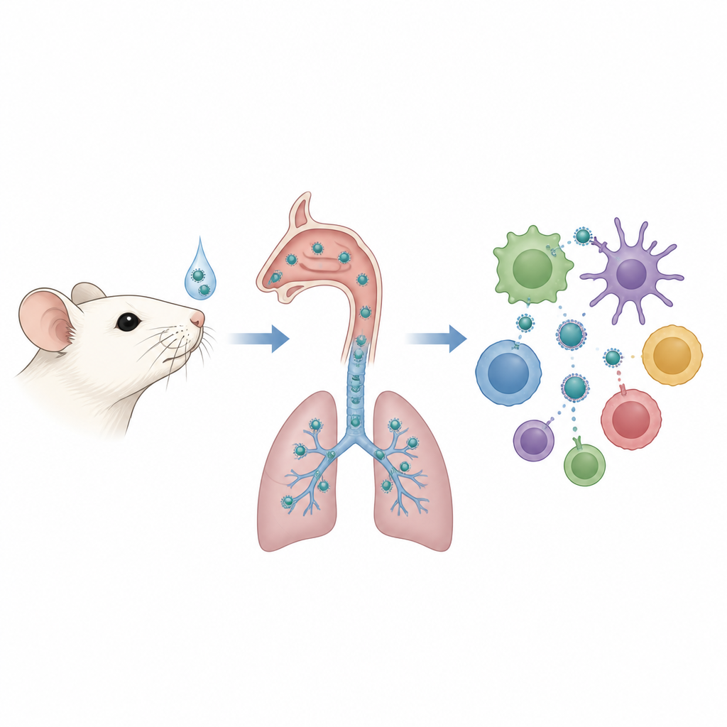
Following the vaccine from nose to lungs
After the vaccine was dripped into the noses of mice, sensitive cameras tracked its glow from one hour to three days later. The light showed that most particles stayed in the nasal passages for up to two days, with some moving down into the lower airways. By about 72 hours, the signal had largely disappeared, suggesting the immune system had cleared or absorbed the particles. At the same time, microscope images and cell counts revealed a rapid surge of immune cells into the nasal tissues, peaking around 24 hours and then fading as the OMVs were removed.
Early foot soldiers rush to the scene
Using detailed flow cytometry, the researchers mapped many types of white blood cells across the nose, lungs, nearby lymph nodes, and spleen. In the nose, there was a strong early wave of neutrophils and inflammatory monocytes, two front line cell types that sense danger and help clean up foreign material. Some less familiar cells called myeloid derived suppressor cells also appeared mainly in the nasal tissues, hinting at local mechanisms that may prevent excessive damage. In the lungs, other sentinels such as alveolar macrophages and specialized dendritic cells took up the OMVs, pointing to a division of labor between upper and lower airways.
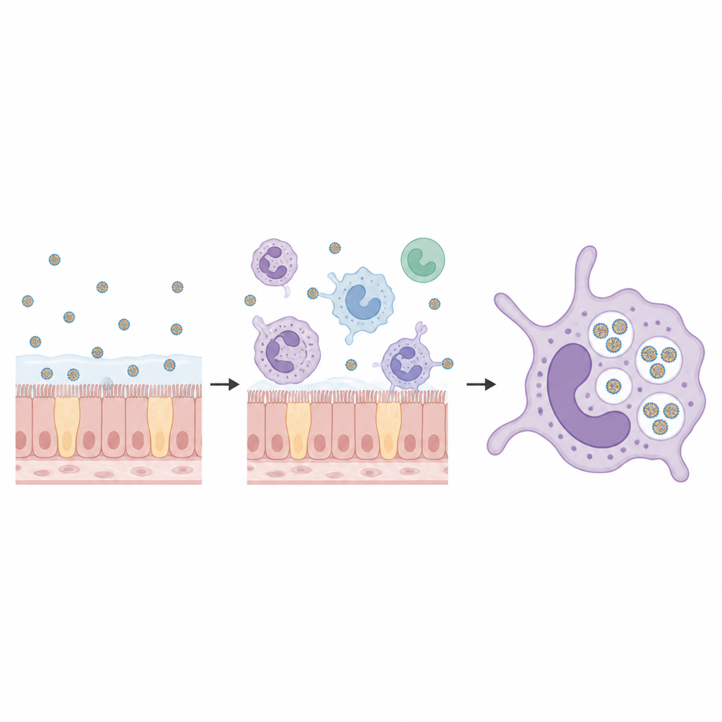
From first response to immune memory
Beyond counting cells, the team looked at activation switches on their surfaces. Many myeloid cells in the nose turned on markers that signal readiness to present antigens and talk to T cells. One striking finding was that a subset of neutrophils in the nasal tissue began to display a molecule usually linked to professional antigen presenting cells. High resolution three dimensional imaging confirmed that these neutrophils physically wrapped around OMV particles and pulled them into internal pockets. In parallel, T cells in the nose quickly showed signs of activation and began to shift into memory like states, followed later by similar changes in the lungs, lymph nodes, and spleen.
What this means for future nose spray vaccines
Altogether, the study shows that this OMV based nose spray vaccine lingers in the nasal passages long enough to spark a fast but controlled immune reaction. Neutrophils and related cells rush in, take up the particles, and switch on features that can help kick start T cell responses and the formation of immune memory. These early events provide a roadmap for how such vaccines may protect against pneumonia causing bacteria and offer clues for designing next generation intranasal vaccines that are both effective and gentle on delicate airway tissues.
Citation: Kanwal, S., To, S.V., Uijen, R. et al. Tracking spatio-temporal dynamics of early immune responses to an intranasal OMV-based pneumococcal vaccine candidate in mice. npj Vaccines 11, 105 (2026). https://doi.org/10.1038/s41541-026-01430-y
Keywords: intranasal vaccine, outer membrane vesicles, pneumococcal immunity, mucosal immune response, neutrophil activation