Clear Sky Science · en
CellCousin2: an optimized system for partial ablation and tracing of regenerative lineages
How the Liver Finds a Way to Heal
Our bodies have a surprising talent for repair, and the liver is one of the best examples. Yet even in this naturally resilient organ, scientists struggle to see exactly which cells step in when damage occurs. This study introduces an improved way to watch liver cells in living zebrafish as some cells are removed and others take over, revealing how different cell families share the work of rebuilding tissue.
Why the Liver’s Backup Plans Matter
The liver can recover from injury using several routes: surviving liver cells can divide, or cells from nearby bile ducts can switch identity and become liver cells. Understanding how these routes are chosen could help explain why some livers recover while others slide into chronic disease. Zebrafish offer a convenient window into this process because their organs are transparent in young animals and they regenerate well. But to untangle who-does-what during healing, researchers need a way to label and remove specific cell groups with great precision.
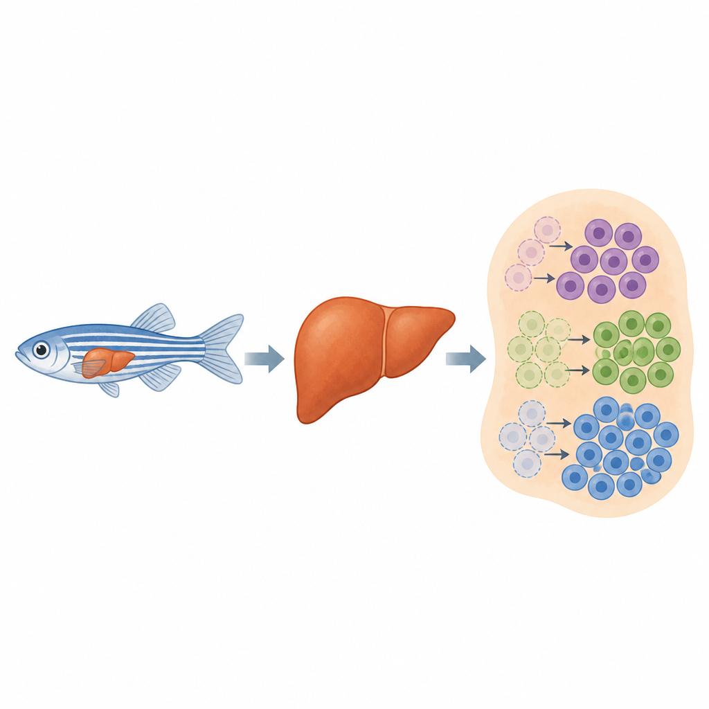
A Color-Coded Map of Liver Cell Families
The team had previously built a tool called CellCousin that uses fluorescent colors to mark liver cells in zebrafish. A genetic switch randomly assigns each liver cell one of several bright colors, so that some can later be killed on demand while others are spared. Watching which colors reappear after injury shows whether healing came from surviving cells or from newcomers. However, the first version suffered from two problems: the genetic switch could flip on its own over time, blurring the history of cell families, and the drug used to kill marked cells had to be delivered at high doses that disturbed the liver even in cells that were not targeted.
Tightening the Genetic Switch
To solve the first problem, the researchers redesigned the genetic switch that controls when cells change color. They fused the common Cre switch to a small “self-destruct” tag that sends it to the cell’s protein disposal system unless two drugs are present at the same time. One drug stabilizes the protein, and the other allows it to enter the nucleus where it can flip the color code. In zebrafish livers, this new dual-control switch stayed almost completely silent without treatment, yet when both drugs were added together during a short window in early life, more than ninety percent of liver cells changed color in a controlled way. This allowed the team to label large cell populations at a chosen time while keeping background noise extremely low as the animals aged.
Gentler Removal of Target Cells
The second improvement focused on safely removing marked cells. The original system used a bacterial enzyme that converts the drug metronidazole into a toxic compound inside labeled cells, but only when the drug was given at high concentrations that also stressed the liver more broadly. The authors swapped this enzyme for a newer version that works much more efficiently. They showed that with this upgrade, they could wipe out the targeted liver cells using one tenth of the previous drug dose. At this lower level, the overall pattern of gene activity in the liver remained close to normal, meaning the tissue’s response mainly reflected the loss of chosen cells rather than side effects of the drug.
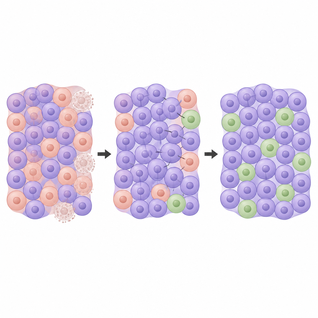
Watching New Liver Cells Take Over
With both upgrades in place, the complete CellCousin2 system could mark different liver cell lineages, remove one subset, and then follow the surviving and newly formed cells for months. After partial removal, some labeled cells clearly survived, while other regions of the liver filled in with cells carrying the default color that had not been labeled before. This pattern matches a mix of self-duplication by spared liver cells and appearance of de novo cells that likely came from other sources such as bile duct cells. Because the color codes are stable over time, researchers can now sort these different groups and compare their properties long after the initial injury.
What This Means for Future Regeneration Research
To a non-specialist, CellCousin2 can be thought of as a highly precise “tag and track” system that marks liver cells in different shades, selectively erases one shade, and then watches how the remaining colors repaint the organ. By making the switch more reliable and the cell removal gentler, this work gives scientists a clearer view of how different cell families cooperate to rebuild damaged liver tissue. The same strategy can be adapted to other organs, offering a powerful way to study how our bodies repair themselves and why some repairs succeed while others fail.
Citation: Hovhannisyan, G.G., Akhourbi, T., Eski, S.E. et al. CellCousin2: an optimized system for partial ablation and tracing of regenerative lineages. npj Regen Med 11, 23 (2026). https://doi.org/10.1038/s41536-026-00473-y
Keywords: liver regeneration, zebrafish model, lineage tracing, cell ablation, cell plasticity