Clear Sky Science · en
Seeing through collagen: integrative pro-regenerative corneal implants for clearer future
Why Clear Windows to the Eye Matter
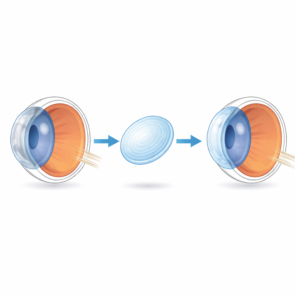
The cornea is the eye’s front window. When it turns cloudy from injury, infection, or disease, vision can drop to legal blindness or worse. Millions of people worldwide need corneal transplants, but suitable donor tissue is scarce, especially in low‑income regions. This article explores how scientists are turning collagen, the body’s own structural protein, into lab‑made corneal implants that could one day replace donor grafts and restore sight more safely and reliably.
From Donor Shortage to Engineered Replacements
Today, full or partial corneal transplantation using donor tissue is the standard treatment for severe corneal disease. While procedures such as penetrating keratoplasty and lamellar keratoplasty can be very successful, they depend on a steady stream of donated corneas and carry risks like rejection, infection, and long healing times. Fully artificial devices, such as early plastic-based corneal prostheses, have helped a few high‑risk patients but often suffer from poor long‑term clarity, inflammation, mechanical failure, or the need for donor tissue as a carrier. The review argues that to treat corneal blindness on a global scale, we need implants that behave more like living tissue yet can be manufactured in large numbers.
Collagen as Nature’s Building Block
Collagen is the main structural protein in the cornea and makes up most of its middle layer, the stroma. There, collagen fibrils are arranged in remarkably uniform, crisscrossing sheets that both strengthen the eye and let light pass almost unhindered. Because collagen is abundant, relatively inexpensive, and already familiar to the body, it is an attractive base material for artificial corneas. However, when collagen is removed from tissue and turned into soft gels, it loses the finely tuned architecture and strong chemical links found in the native cornea. On its own, it tears easily, swells, and can be digested by enzymes in diseased or inflamed eyes. The central challenge is to recreate, in a lab, a collagen network that is strong, transparent, and stable enough for everyday life while still welcoming the patient’s own cells and nerves.
Designing Strong, Clear Collagen Scaffolds
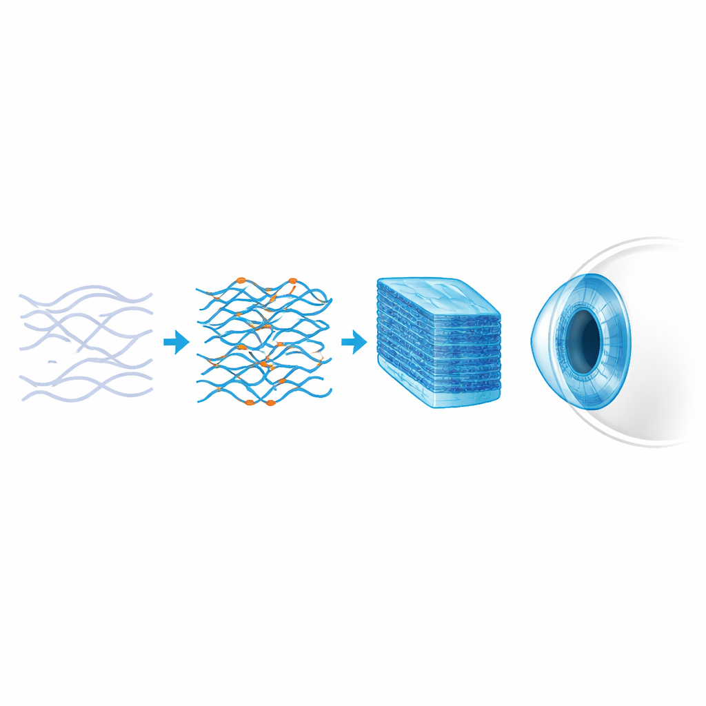
Researchers have developed a toolbox of strategies to toughen and organize collagen without sacrificing clarity. Physical methods compress or guide the self‑assembly of collagen fibrils to form denser, lamellar structures that resemble natural stroma. Chemical “crosslinkers” act like molecular staples, tying collagen strands together so they resist tearing and enzymatic breakdown. Some are simple treatments with light and vitamin-like molecules; others use small reactive chemicals or flexible polymers such as PEG to form denser, more elastic networks. Interpenetrating polymer networks go a step further by weaving a second, supportive polymer through the collagen, boosting strength, suture‑holding ability, and resistance to clouding, while preserving a watery, cell‑friendly interior. Emerging fabrication methods—casting, electrospinning of fine fibers, and 3D bioprinting—allow researchers to shape these materials into curved, layered structures that better match the eye’s natural geometry and guide cell alignment.
Beyond Structure: Guiding Healing and Delivering Drugs
Modern collagen implants are not just passive lenses; they can be tuned to actively steer healing and fight complications. Surface patterns with microscopic grooves or ridges encourage corneal cells and stem cells to align and lay down new, orderly collagen, reducing scar formation. Nanofibers and microfibers embedded in the gel help distribute surgical forces so stitches do not rip through the material. Antibiotics and anti‑inflammatory drugs can be built into the collagen network or attached to its surface for slow, local release, lowering the risk of infection and rejection after surgery. Some designs even incorporate nanoparticles so that doctors can monitor implant position and tissue response by advanced imaging, turning the graft into a combined therapeutic and diagnostic device.
Early Human Trials and the Road Ahead
Several collagen-based corneal implants have already reached animal studies and early clinical trials. Implants made from recombinant or animal‑derived collagen, strengthened by carefully chosen crosslinkers, have been sutured or slid into diseased corneas. Over months and years of follow‑up, many have stayed clear, become populated by the patient’s own cells, and regained nerve connections and sensitivity, often without long‑term immune‑suppressing drugs. Newer versions use stronger double‑crosslinked pig collagen and minimally invasive, suture‑free surgery, showing promising improvements in corneal thickness, shape, and vision in people with advanced disease such as keratoconus. The authors conclude that while challenges remain—especially fully matching the native cornea’s mechanical toughness and proving long‑term safety at scale—collagen-based artificial corneas are rapidly evolving from experimental constructs into realistic, off‑the‑shelf alternatives to donor tissue, with the potential to open a clearer future for millions at risk of corneal blindness.
Citation: Huang, X., Islam, M.M., Watson, S.L. et al. Seeing through collagen: integrative pro-regenerative corneal implants for clearer future. npj Regen Med 11, 21 (2026). https://doi.org/10.1038/s41536-026-00471-0
Keywords: corneal implants, collagen biomaterials, artificial cornea, tissue engineering, vision restoration