Clear Sky Science · en
O-GlcNAcylation regulates microglial neuroinflammation in Parkinson’s disease
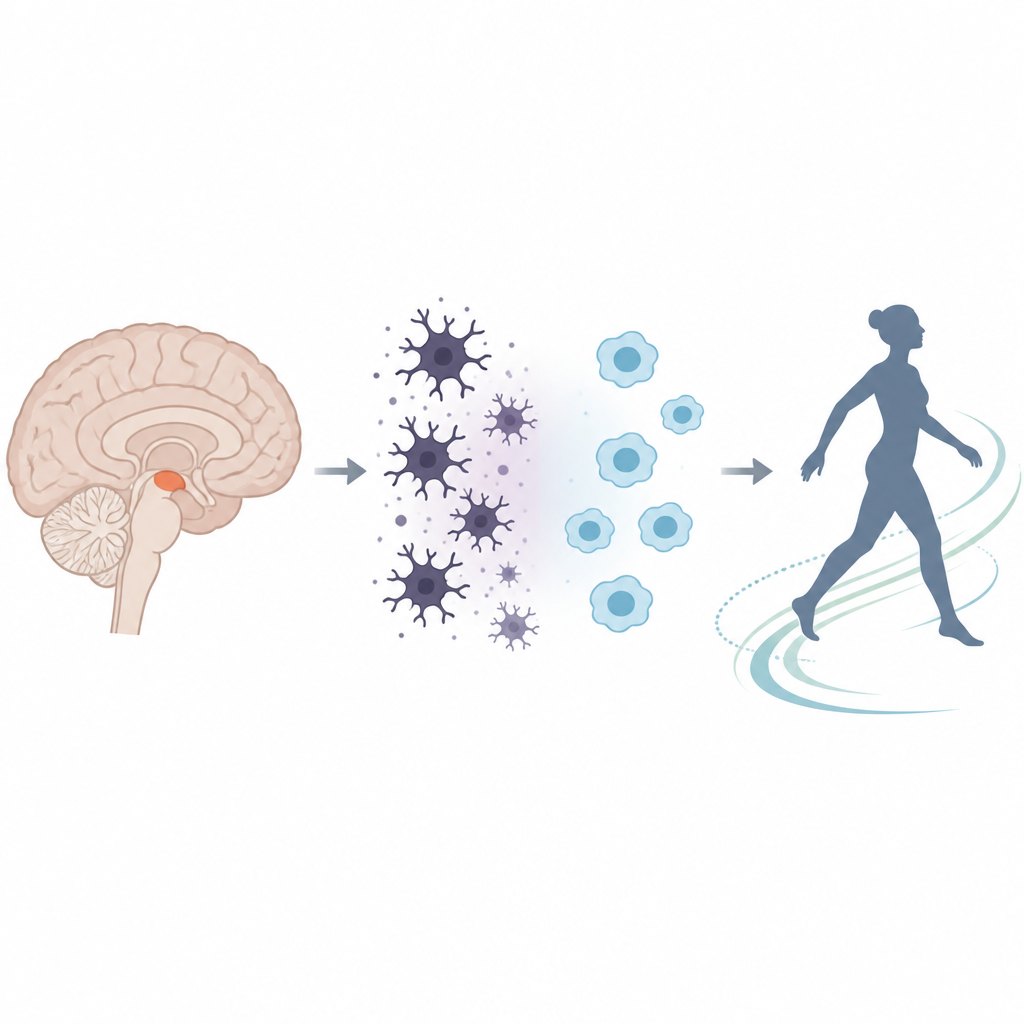
Why brain immune cells matter in Parkinson’s
Parkinson’s disease is best known for its shaking hands and slowed movements, but deep inside the brain another story is unfolding. Tiny immune cells called microglia can either protect nerve cells or help drive their damage. This study explores how a sugar-based chemical tag inside these cells helps decide which path they take, and whether gently tweaking that tag can calm harmful brain inflammation.
A chemical switch inside brain guardians
Microglia patrol the brain, clearing debris and responding to injury. They can shift between a calm, caretaking mode and an aggressive, inflammatory mode. The authors focused on a subtle chemical mark called O-GlcNAcylation, a small sugar attached to many proteins inside cells and influenced by nutrients. They asked whether low levels of this mark in microglia are linked to the chronic brain inflammation seen in Parkinson’s disease, and whether restoring it might nudge microglia back toward a more protective state.
Clues from human brains and mouse models
Examining brain tissue from people who had Parkinson’s, the researchers found that the affected region called the substantia nigra not only had the expected loss of dopamine-producing neurons and buildup of alpha-synuclein protein, but also showed strikingly reduced O-GlcNAcylation. This drop was especially clear in microglia, which at the same time displayed strong signs of activation and inflammation. Proteins that drive inflammatory signaling and a molecular machine called the inflammasome were elevated, and markers of a calm, tissue-supporting microglial state were reduced. Together, these observations tied lower O-GlcNAcylation to a more aggressive immune environment in the Parkinson’s brain.
Testing a calming strategy in mice
To probe cause and effect, the team turned to a mouse model in which a bacterial component, lipopolysaccharide, is injected into the substantia nigra to trigger local inflammation and gradual loss of dopamine neurons. In these mice, movement became clumsy, brain inflammation surged, and O-GlcNAcylation levels fell, echoing the human findings. When the researchers boosted O-GlcNAcylation using two different compounds, glucosamine and Thiamet G, the picture changed. Mice moved more normally, dopamine-producing neurons were preserved, and many signs of inflammation and oxidative stress dropped, including inflammatory enzymes, reactive oxygen molecules, and inflammasome activity.
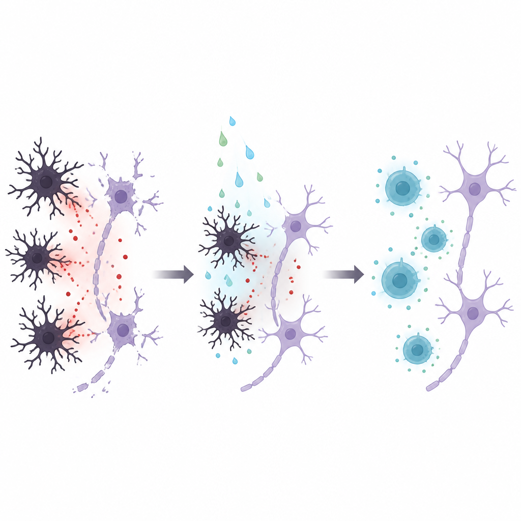
How microglia change their behavior
The scientists next studied purified microglia grown in dishes. When exposed to lipopolysaccharide, these cells ramped up inflammatory genes and released damaging molecules that, in turn, harmed nearby nerve-like cells. Boosting O-GlcNAcylation reversed this pattern: inflammatory markers fell, protective and homeostatic markers rose, and the secretions of microglia became less toxic for neurons. At the molecular level, raising O-GlcNAcylation reduced the movement of key NF-kappa B proteins into the cell nucleus, a step normally needed to switch on many inflammatory genes. The study also showed that dialing O-GlcNAcylation up or down, even without added inflammatory triggers, could tilt microglia toward more helpful or more harmful activation states.
What this means for future treatment ideas
For a lay reader, the main message is that Parkinson’s disease is not only a problem of dying nerve cells but also of stressed brain immune cells stuck in an inflammatory mode. This work suggests that a nutrient-sensitive sugar tag on proteins acts as a kind of internal dimmer switch for microglial behavior. When the tag is low, microglia are more likely to fuel inflammation and nerve-cell loss; when it is restored to balanced levels, they shift toward protecting brain tissue. While the compounds used here are tools rather than ready-made treatments, the study points to O-GlcNAcylation as a promising control point for calming harmful brain inflammation in Parkinson’s and possibly related disorders.
Citation: Kim, D.Y., Kim, SM., Lee, C. et al. O-GlcNAcylation regulates microglial neuroinflammation in Parkinson’s disease. npj Parkinsons Dis. 12, 121 (2026). https://doi.org/10.1038/s41531-026-01319-6
Keywords: Parkinson’s disease, microglia, neuroinflammation, O-GlcNAcylation, dopaminergic neurons