Clear Sky Science · en
HIF-1–regulated TPM3 links hypoxia to motility and invasion beyond the hypoxic fraction in triple-negative breast cancer
Why oxygen-starved tumors matter
Many aggressive breast cancers grow so quickly that parts of the tumor run low on oxygen. This low-oxygen, or hypoxic, environment makes cancers harder to treat and more likely to spread. In triple-negative breast cancer—a form that lacks common drug targets—understanding how hypoxia changes cancer cells could reveal new ways to slow or stop metastasis. This study focuses on a structural protein inside cells, called TPM3, and uncovers how it helps hypoxic tumor regions drive movement and invasion throughout the tumor, even into better-oxygenated areas.
A structural helper gone rogue
TPM3 normally helps organize the inner scaffolding of cells, giving them shape and enabling movement. The researchers first asked whether breast tumors show altered levels of this protein. By mining large patient datasets, they found that TPM3 levels are higher in breast cancer tissue than in normal breast tissue, and even higher in triple-negative tumors. Patients whose tumors carried more TPM3 tended to have poorer overall survival. TPM3 levels also rose in tumors with stronger molecular signatures of hypoxia, hinting that oxygen shortage and this structural protein might be closely linked. 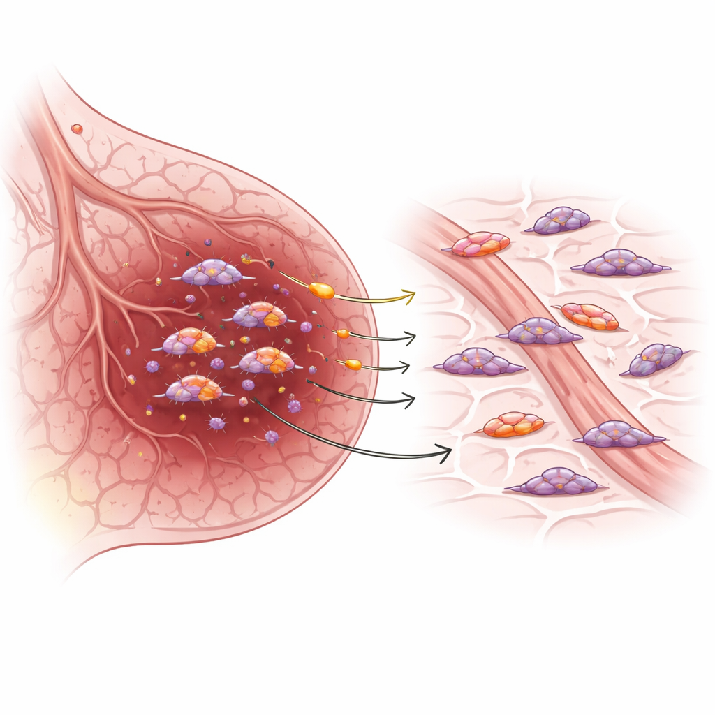
How low oxygen tunes cell movement
To probe this link, the team grew triple-negative breast cancer cells under different oxygen levels, including steady low oxygen, near-complete oxygen deprivation, and cycling between the two. In all these conditions, TPM3 levels went up at both the RNA and protein level. They showed that this rise depends largely on HIF-1, a master switch that turns on many genes when oxygen is scarce. Under hypoxia, TPM3 lined up along actin filaments—the cables that cells use to push and pull themselves forward. When the scientists reduced TPM3 using genetic tools or a small-molecule inhibitor, cells lost their usual rounded shape, developed ragged trailing edges, and formed weaker actin structures at the front. These changes translated into slower migration across a surface and less ability to invade through a gel-like barrier, especially under low oxygen, even though the cells remained alive.
Turning a weakness into a treatment angle
The study then examined how blocking TPM3 might work with current therapies. In lab dishes, inhibiting TPM3 did not make cells more sensitive to radiation, but it did combine especially well with two standard chemotherapies, Doxorubicin and Paclitaxel. Using drug-interaction analyses, the team found strong synergy: together, TPM3 inhibition and these drugs reduced cell viability more than either alone, without obvious added toxicity to basic cell survival in the short term. This suggests that TPM3-targeting drugs, some of which have already shown acceptable tolerance in animal studies, could be paired with existing chemotherapy to better control aggressive tumors by curbing their ability to move and spread. 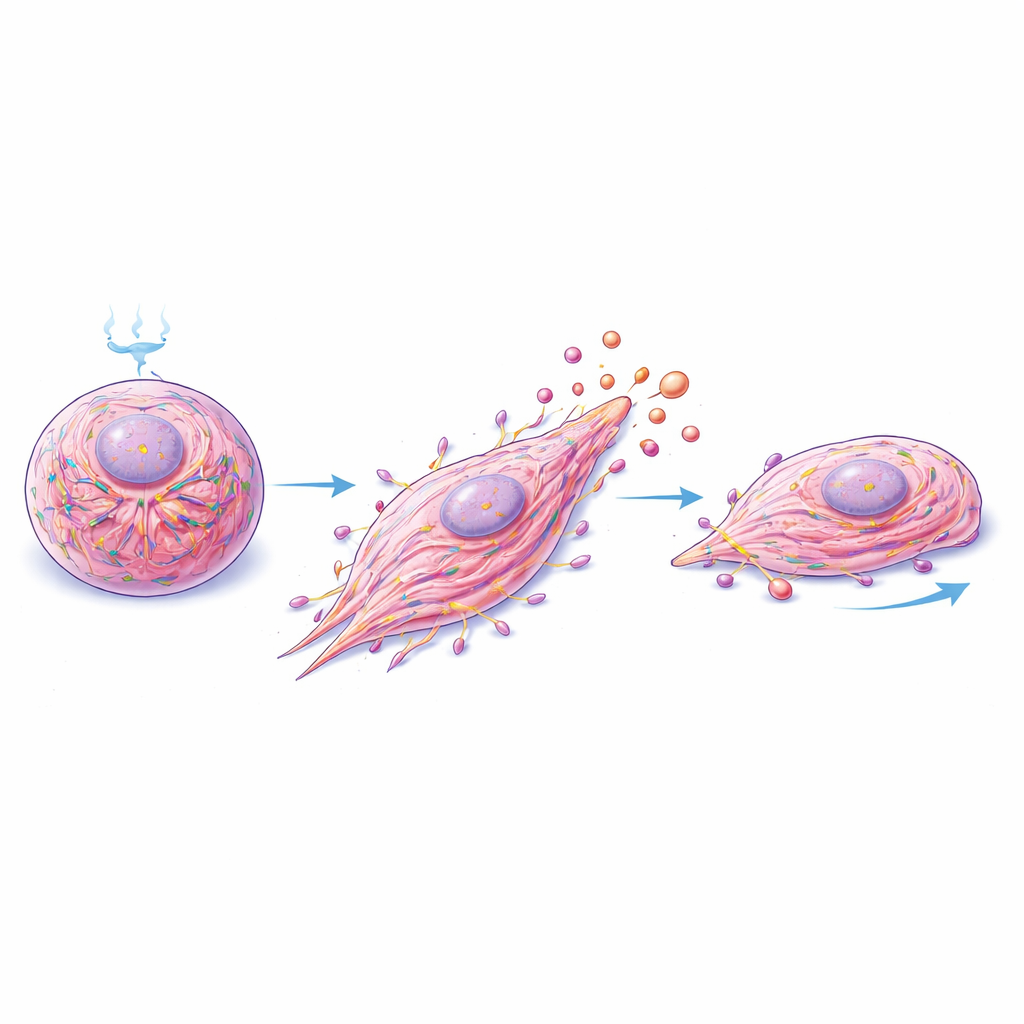
Messages in microscopic parcels
Hypoxic tumor regions do not act in isolation; they communicate with neighboring, better-oxygenated cells. The researchers explored whether TPM3 plays a role in this cross-talk. When they collected the liquid surrounding hypoxic cells and applied it to normoxic cells, the recipient cells moved faster. But if TPM3 had been reduced in the donor cells, this boost in movement disappeared. Blocking uptake of extracellular vesicles—tiny membrane-bound parcels that cells release—also blunted the effect. Electron microscopy and particle tracking showed that hypoxia makes cancer cells release more vesicles of similar size. Crucially, TPM3 itself was detected inside these vesicles, and its levels were higher in vesicles from hypoxic cells, confirming that TPM3 is shipped out as cargo.
What this means for patients
Altogether, the work paints TPM3 as a key middleman connecting low oxygen, cell shape, and cell movement in triple-negative breast cancer. Under hypoxia, HIF-1 boosts TPM3, which stabilizes the internal scaffolding that powers migration and invasion. At the same time, hypoxic cells package TPM3 into extracellular vesicles that can be taken up by nearby oxygenated cells, encouraging those cells to become more motile as well. This means that a relatively small hypoxic fraction of a tumor can influence behavior across the whole mass. By highlighting TPM3 as both a marker of hypoxic adaptation and a druggable driver of motility, the study suggests that targeting this protein—especially in combination with standard chemotherapies—could help limit the spread of triple-negative breast cancer and improve patient outcomes.
Citation: Zhou, C., Crusher, J.T., Friesen, K. et al. HIF-1–regulated TPM3 links hypoxia to motility and invasion beyond the hypoxic fraction in triple-negative breast cancer. npj Breast Cancer 12, 64 (2026). https://doi.org/10.1038/s41523-026-00927-y
Keywords: triple-negative breast cancer, tumor hypoxia, cell motility, extracellular vesicles, TPM3