Clear Sky Science · en
Colouring dysbiosis: FetB-dependent Mn-PPIX produced by Porphyromonas gingivalis shapes the oral microbiota
Why Mouth Bacteria Change Color Matters
Your mouth is home to bustling communities of bacteria that usually live in balance and help keep you healthy. But when that balance is disturbed, these tiny residents can drive gum disease and even contribute to problems elsewhere in the body. This study reveals how one key gum‑disease bacterium, Porphyromonas gingivalis, responds to blood leaking into the gums by making unusual pink, glowing pigments that can kill some neighboring bacteria. By uncovering how these pigments are made and what they do, the researchers shed light on how a healthy mouth community can slide into harmful imbalance—and how we might one day prevent it.
A Color Shift in the Gums
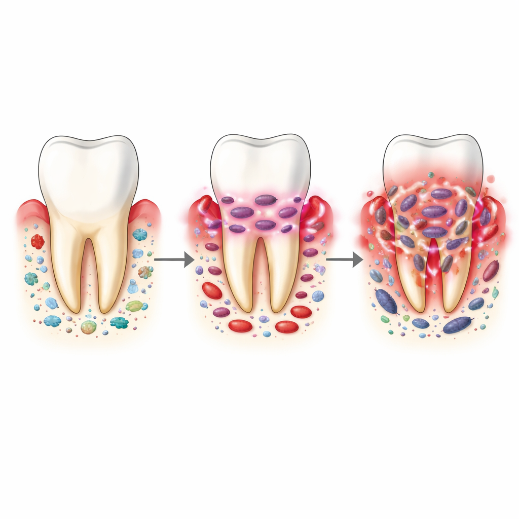
Periodontitis, a common chronic gum disease, arises when normally friendly mouth bacteria shift into a damaging partnership known as dysbiosis. P. gingivalis is considered a “keystone” member of this harmful group: even in modest numbers it can reshape the entire community and nudge the immune system toward long‑lasting inflammation. This organism cannot make its own building blocks called porphyrins, so it scavenges them from haemoglobin, the red pigment in blood. In a healthy mouth, free haemoglobin is scarce; in diseased gums, bleeding raises its levels—but still not to the very high concentrations traditionally used in lab studies. The researchers therefore grew P. gingivalis under haemoglobin levels that mimic real gum conditions, from low to high, and watched how its color and behavior changed.
Discovery of a Pink, Glowing State
Under very high haemoglobin, P. gingivalis produced the familiar black pigmentation seen in lab cultures. At very low haemoglobin, its cells appeared pale. Strikingly, at intermediate levels—similar to those in the crevice between tooth and gum during early and progressing disease—the bacteria turned a distinctive pink and became strongly fluorescent under ultraviolet light. Chemical analysis of pigments extracted from these pink cells showed a mixture of known and novel porphyrins: regular haem, protoporphyrin IX (PPIX), manganese‑substituted PPIX (Mn‑PPIX), and at least one related compound. These findings revealed that the bacterium does not just passively collect haem from its surroundings; it actively remodels these molecules, especially at the very stages when the gum environment is changing.
The Enzyme Behind the Pigment Trick
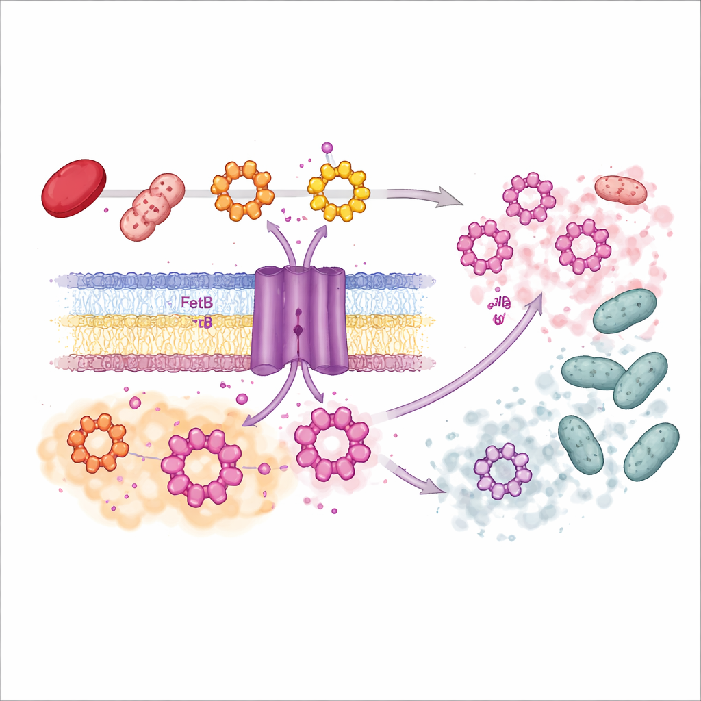
To understand how P. gingivalis swaps metals into porphyrins, the team searched its genome for relatives of enzymes that insert metal ions into ring‑shaped molecules. They homed in on a protein called FetB, already known to bind haem. Using structural biology, they solved FetB’s three‑dimensional shape at atomic detail and found it closely resembled known metal‑inserting enzymes. In test‑tube experiments, purified FetB readily inserted manganese and cobalt—but not iron—into a porphyrin‑like ring, confirming its role as a metal‑inserting catalyst. When the scientists deleted the fetB gene from P. gingivalis, production of Mn‑PPIX dropped sharply, and the fluorescent signal was greatly reduced. Restoring the gene brought Mn‑PPIX back, showing FetB is a major driver of this pink pigment pathway.
How Pink Pigment Rewrites the Neighborhood
The team then asked what Mn‑PPIX does to other mouth microbes. In a simple plate assay, they placed purified Mn‑PPIX near lawns of different oral bacteria and looked for clear zones where growth was blocked. Mn‑PPIX strongly inhibited several common commensals, including Streptococcus mitis, Streptococcus salivarius, Enterococcus faecalis, and oral Lactobacillus species, even at relatively low concentrations. Other species, such as Streptococcus oralis, S. gordonii, and S. mutans, were unaffected. This selective action means the pigment can act like a targeted weapon: it weakens some early, health‑associated colonizers while sparing others and the producer itself. Because Mn‑PPIX tends to bind to the surface of P. gingivalis rather than diffuse freely, its effects are likely concentrated in the immediate biofilm neighborhood, where bacteria cluster tightly on tooth and gum surfaces.
From Colored Plaques to New Treatment Ideas
Together, these findings suggest that as gums start to bleed and leak haemoglobin, P. gingivalis senses this nutrient cue and rewires its porphyrin chemistry through FetB, creating manganese‑rich, pink pigments on its surface. These pigments, in turn, selectively suppress certain beneficial or neutral neighbors, tilting the community toward a dysbiotic, disease‑favoring state and helping sustain chronic inflammation that supplies more nutrients. Understanding this chain of events opens fresh avenues for therapy: limiting haemoglobin leakage through better control of early inflammation, blocking FetB or Mn‑PPIX formation, or scavenging these extracellular pigments might help restore a healthier balance of mouth microbes and slow or prevent gum disease progression.
Citation: Phonok, Y., Pyne, A., Liu, S. et al. Colouring dysbiosis: FetB-dependent Mn-PPIX produced by Porphyromonas gingivalis shapes the oral microbiota. npj Biofilms Microbiomes 12, 76 (2026). https://doi.org/10.1038/s41522-026-00942-8
Keywords: oral microbiome, gum disease, Porphyromonas gingivalis, bacterial pigments, dysbiosis