Clear Sky Science · en
Over four minutes of pyruvate T1 using chemically and physically induced deceleration of relaxation
Why slowing a fading signal matters
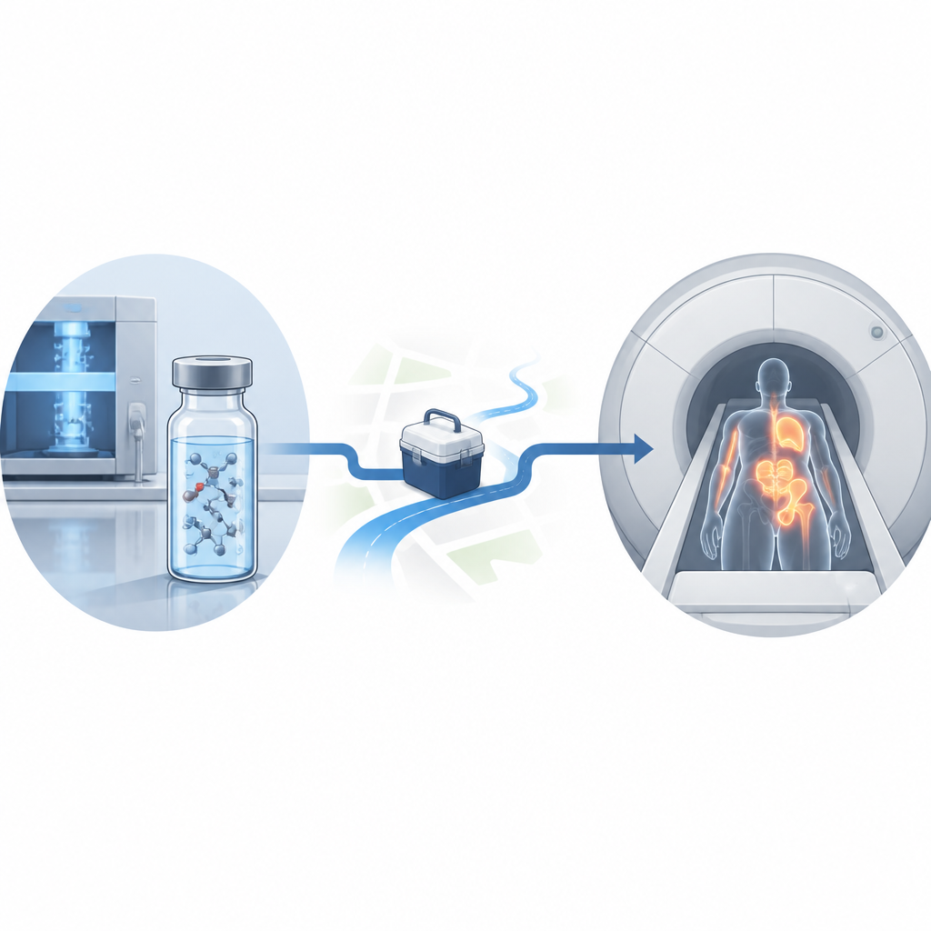
Medical scanners that track the body’s chemistry in real time rely on signals that fade in under a minute. This study shows how to keep one of the most important of those signals alive for over four minutes. By fine tuning the recipe and handling of a small molecule called pyruvate, the researchers greatly extend how long its magnetic signal lasts, which could make metabolic MRI scans clearer, more reliable, and easier to perform in both research and clinical settings.
A chemical spotlight on living tissue
Hyperpolarized metabolic MRI is a technique that briefly turns certain molecules into powerful beacons inside the body. One of the workhorses of this approach is [1-13C]pyruvate, a labeled version of a natural fuel molecule that cells rapidly convert into other substances. When injected, its bright but short lived signal lets doctors and scientists watch tumors, heart muscle, or inflamed tissue process energy in real time. However, the signal begins to fade as soon as pyruvate is hyperpolarized, and it often weakens substantially during the steps needed for quality checks, transport from the polarizer to the scanner, and injection into a patient or sample.
Looking for lost signal
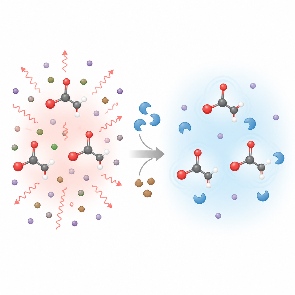
The team set out to understand exactly why pyruvate’s signal disappears and how to slow that loss. They measured how long the signal lasted at magnetic fields ranging from millionths of a tesla up to 9.4 tesla while systematically changing the pyruvate solution. They tested different solvents, buffers, and additives, removed dissolved oxygen, and even replaced some of pyruvate’s hydrogen atoms with heavier deuterium. More than 4300 measurements and computer simulations helped them tease apart which interactions at the atomic level do the most damage to the signal in different magnetic environments.
Building a calmer environment for pyruvate
The researchers found that no single trick was enough; instead, many small changes added up. Using heavy water rather than regular water reduced certain magnetic interactions with surrounding hydrogen atoms. Adding common biocompatible helpers such as Tris buffer and the chelating agent EDTA kept metal ions and other paramagnetic impurities away from pyruvate, echoing a recently described “chemical shield” effect. Removing dissolved oxygen further reduced harmful interactions, especially at low magnetic fields where transport losses matter most. Finally, swapping pyruvate’s methyl hydrogens for deuterium gave an extra boost at lower fields. Together, these changes stretched the key relaxation time, called T1, from roughly half a minute to more than four minutes under ideal conditions.
From test tube to living cells
To see whether these gains matter in practice, the team applied an optimized recipe in cell experiments. They used hyperpolarized pyruvate to monitor lactate production in HeLa cancer cells, comparing a standard solution with improved versions prepared in heavy water, with or without a radical filtration step. Even without changing the biology, just altering the solution roughly doubled the lifetime of the signal at low field and more than doubled the strength of the lactate signal detected in the cells. In realistic transfer times similar to those in clinical workflows, the optimized mixture preserved far more of the initial polarization, directly translating into higher signal to noise ratio.
What this means for future scans
For non specialists, the key message is that careful chemistry can buy valuable extra minutes for a delicate signal that underpins advanced MRI scans of metabolism. By reducing many small sources of magnetic disturbance in the pyruvate solution, the researchers show that it is possible to transport and use hyperpolarized pyruvate with far less loss of signal. This could let clinicians image smaller tumors, follow treatment responses more precisely, or extend measurement windows without changing anything in the patient, only in the contrast agent they receive.
Citation: Peters, J.P., Teleanu, F., Zou, H. et al. Over four minutes of pyruvate T1 using chemically and physically induced deceleration of relaxation. Nat Commun 17, 4561 (2026). https://doi.org/10.1038/s41467-026-73214-w
Keywords: hyperpolarized MRI, pyruvate imaging, nuclear spin relaxation, magnetic resonance contrast, metabolic imaging