Clear Sky Science · en
Smart event-triggered MINFLUX microscopy to catch and follow rare events
Watching Cells Only When Something Happens
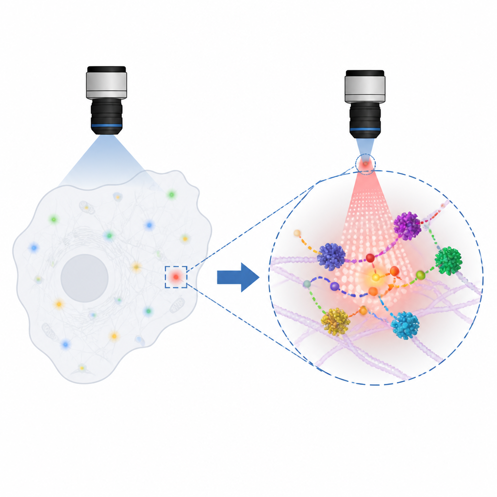
Our cells are full of brief, tiny events that are easy to miss, like a bubble budding from the cell surface or a virus starting to form. This study introduces a way for a powerful microscope to “pay attention” only when such rare events occur, so researchers can zoom in at the exact right moment and see details just a few billionths of a meter across.
A Smarter Kind of Super-Resolution Microscope
The work builds on MINFLUX, a cutting-edge microscope that can pinpoint single fluorescent molecules with nanometer precision and track them on microsecond timescales. The drawback of MINFLUX is that it normally watches just one molecule at a time, making experiments slow and difficult to use on living cells where many things happen quickly and at once. The authors solved this by creating event-triggered MINFLUX, or etMINFLUX, which combines a fast, lower-resolution confocal view of a large cell area with the ultra-detailed MINFLUX zoom. A computer continuously analyzes the confocal images in real time and, whenever it spots a pre-defined change in the cell, automatically switches the microscope into MINFLUX mode at that tiny region.
How the Smart System Works
In practice, etMINFLUX scans regions as large as tens of micrometers using gentle confocal light and runs custom analysis programs written in Python. These programs look for patterns such as bright spots appearing, growing, or staying put, which can signal a structure of interest. As soon as an event is detected, the system pauses the confocal scan and redirects the MINFLUX probe to a very small region, often about a micrometer across or less. Because this region is tiny, MINFLUX can quickly collect many precise molecular tracks there, making better use of its time and of the light hitting the cell. Once the detailed measurement is done, the microscope automatically returns to scanning and waiting for the next event, allowing long, unattended experiments.
Following Lipids, Endocytic Bubbles, and Budding Viruses
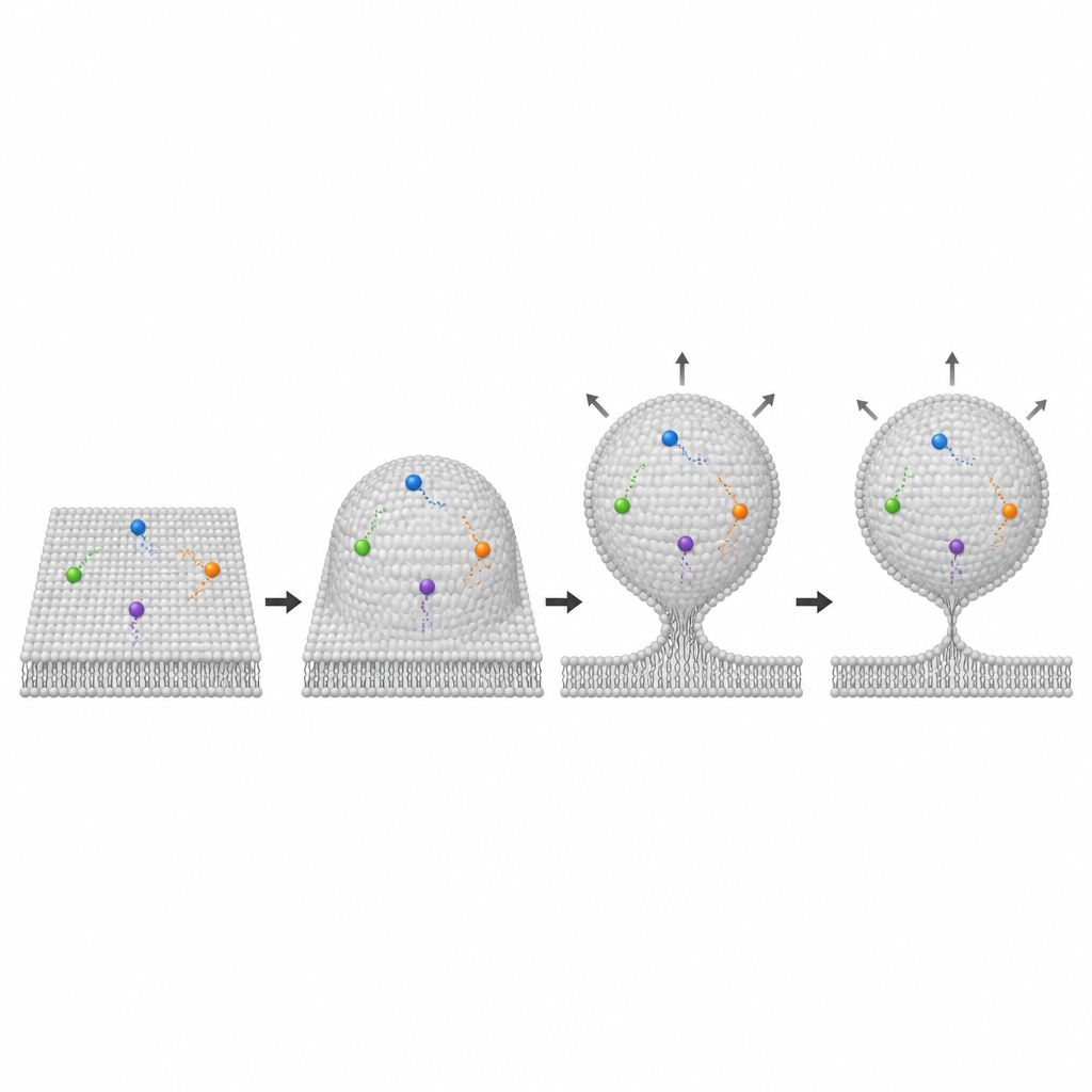
To show what etMINFLUX can do, the team applied it to three different cellular situations. First, they looked at caveolae, small pockets in the cell’s outer membrane associated with signaling, fat handling, and disease. By detecting bright clusters of a caveola marker, the system could rapidly trigger MINFLUX tracking of dye-labeled lipids within these pockets. From hundreds of such regions, the researchers found that one lipid type, related to sphingomyelin, diffused more slowly and appeared more enriched inside caveolae than another, suggesting these pockets selectively shape how certain lipids move. Second, they targeted rare and fast events where the membrane folds inward to form endocytic bubbles. By detecting build-up of a protein called dynamin, which helps pinch off these bubbles, etMINFLUX captured three-dimensional outlines of budding vesicles in living cells. It measured features like bubble size and the length of the narrow neck connecting them to the surface, reaching nanometer-level agreement with earlier electron microscopy but now in a living, moving cell.
Watching Virus Budding Sites Over Minutes
The third test focused on assembly sites of HIV-1, tracked through clusters of a structural protein called Gag. These sites form and evolve over many minutes, so the authors used etMINFLUX to follow the same spots repeatedly with alternating confocal and MINFLUX recordings. The system detected slowly growing Gag-rich regions and measured how a cholesterol-based probe diffused in the surrounding membrane. Surprisingly, most sites showed only modest changes in membrane fluidity and often remained fairly flat, while only a minority developed clear bulges or budding shapes suggestive of forming virus particles. The approach also revealed how hard it would be to reliably catch such slow, subtle changes by hand, showing that automated, event-driven control is crucial for building coherent timelines across many cells.
Why This Matters for Studying Living Cells
Overall, event-triggered MINFLUX turns an extremely precise but slow microscope into a far more efficient and practical tool for live-cell studies. By letting the instrument decide when and where to zoom in, it reduces wasted recording time, increases the fraction of useful data by several fold, and limits unnecessary light exposure that can harm cells. This makes it possible to map the shapes and motions of tiny membrane structures and potential virus budding sites in three dimensions and in real time, opening the door to studying many fast or rare processes in living cells that were previously out of reach.
Citation: Alvelid, J., Koerfer, A. & Eggeling, C. Smart event-triggered MINFLUX microscopy to catch and follow rare events. Nat Commun 17, 4558 (2026). https://doi.org/10.1038/s41467-026-73176-z
Keywords: super resolution microscopy, MINFLUX, live cell imaging, membrane dynamics, virus budding