Clear Sky Science · en
A modular multi-color fluorescence microscope for simultaneous tracking of cellular activity and behavior
Watching tiny animals in action
How do muscles and brain cells work together while an animal explores its world? For creatures that are only a few millimeters long, this question has been hard to answer, because getting close enough to see cells usually means giving up a wide view of behavior. This article introduces a simple, affordable microscope that lets scientists and students watch both cellular activity and full-body movement in tiny animals at the same time.
A simple tool built from ready-made parts
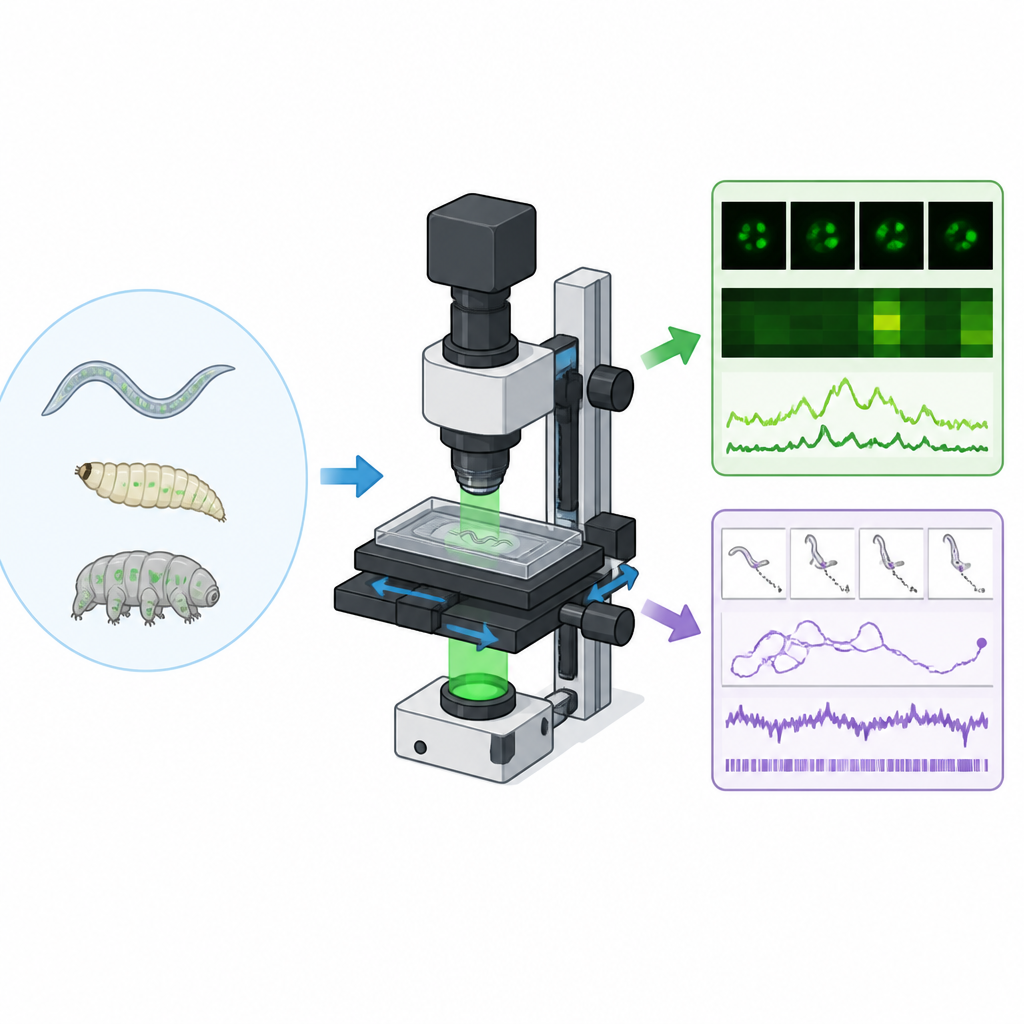
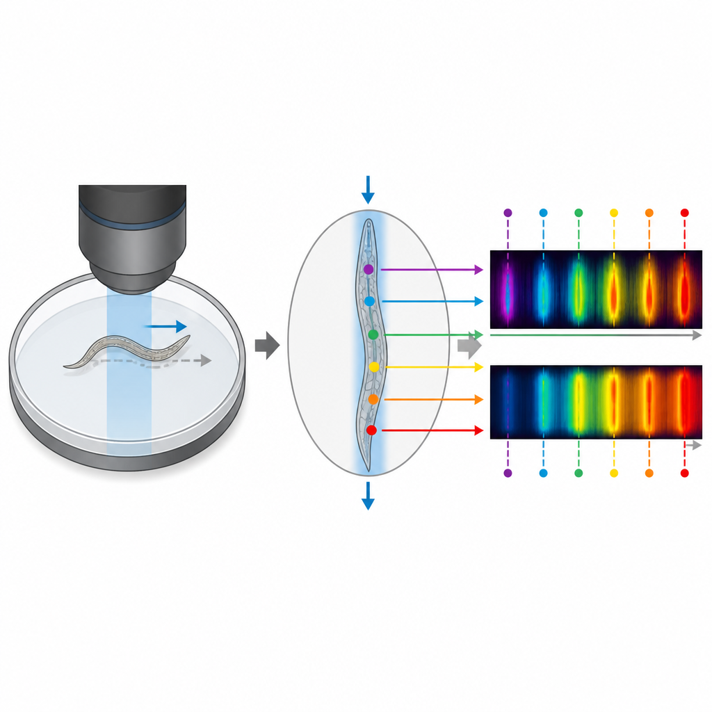
The authors describe a new fluorescence tracking microscope that is assembled entirely from standard commercial components in about three hours. Instead of moving the animal around under a fixed lens, the whole microscope rides on a motorized stage while the animal’s arena stays still. This design reduces shaking and makes it easy to add extras such as temperature control or light sources for stimulation. By swapping a few optical pieces, the same system can switch between brightfield, single-color, and dual-color fluorescence modes, and can zoom from whole animals down to individual neurons.
Software that follows the animal on its own
To control the hardware, the team built a cross-platform program called GlowTracker. This software reads images from the camera, moves the stage to keep the animal in view, and performs basic tracking in real time. It can automatically measure pixel size and camera orientation, align different color channels, and run simple scripted routines. Tests showed that the system can follow animals over large plates for tens of minutes to hours, with frame rates high enough to catch fast changes in muscle and nerve activity.
Linking movement and muscle activity
Using fruit fly larvae that crawl using waves of muscle contraction, the researchers showed how the microscope connects posture, odor-guided navigation, and muscle activity. Larvae crawled across a plate containing a gradient of vinegar scent while their muscles glowed in two colors, one reporting calcium levels and one providing a stable reference. The tracking kept each larva centered even as it roamed centimeters across the arena. From the dual-color movies, the team reconstructed body shape and extracted patterns of muscle activation along the body, revealing how peristaltic waves support straight crawling and turning, and allowing comparisons between larvae that successfully reached the odor source and those that did not.
Peeking into single neurons and predator prey games
The same setup, adjusted for higher magnification, was used to monitor individual touch-sensitive neurons in freely moving roundworms. When the plate was gently vibrated, a specific neuron in the tail showed a calcium signal that rose and fell in distinct ways depending on whether the worm fled forward or reversed direction, demonstrating that the system can capture subtle neural dynamics in motion. In another set of experiments, the dual-color mode followed red-labeled predatory nematodes hunting green-labeled prey. The movies captured full chase sequences, and the green signal around the predator’s mouth spiked whenever it bit and fed, confirming predictions from earlier behavior models about when real contact occurs.
Tracking long journeys and unlabeled animals
The modular design also enables hour-long recordings with minimal fading of fluorescence. In one example, the microscope followed individual worms as they foraged, recording feeding muscle activity, reversals, and speed over large distances. In brightfield mode, the same hardware tracked tardigrades, tiny “water bears” that are not genetically labeled. By combining tracking with pose estimation software, the authors analyzed leg movements over many minutes, uncovering different gait patterns and how they relate to turning and walking speed.
Why this matters for science and teaching
In conclusion, the work shows that careful design and modern camera technology make it possible to build a low-cost, easy-to-use microscope that connects what cells are doing with how whole animals behave. Because it relies on off-the-shelf parts and open-source software, laboratories and classrooms without access to custom engineering can now study complex behaviors, ecological interactions, and gait patterns in tiny animals while still seeing the underlying cellular activity.
Citation: Ramahefarivo, E., Böger, L., Saichol, T. et al. A modular multi-color fluorescence microscope for simultaneous tracking of cellular activity and behavior. Nat Commun 17, 4412 (2026). https://doi.org/10.1038/s41467-026-72710-3
Keywords: fluorescence microscopy, behavior tracking, calcium imaging, C. elegans, Drosophila larvae