Clear Sky Science · en
An epifluorescence microscope design for naturalistic behavior and cellular activity in freely moving Caenorhabditis elegans
Watching tiny worms live their normal lives
To understand how brains control behavior, scientists need to see what nerve cells are doing while an animal moves naturally, not just when it is pinned down under a heavy microscope. This study introduces Wormspy, a simple, low-cost microscope and software system that lets researchers watch both the movements and the glowing activity of cells inside tiny roundworms as they roam freely, offering a window into how nervous systems work in real time.
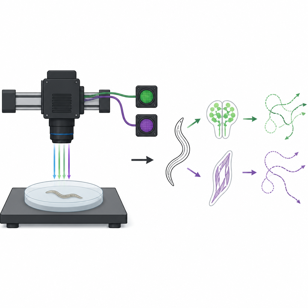
A small animal with a big role in brain research
The work centers on Caenorhabditis elegans, a millimeter-long soil worm that has become a favorite in biology labs. These worms are nearly see-through and have a fixed set of cells, making it possible to follow individual muscles and neurons across different animals. By engineering the worms so that specific cells glow when calcium levels change, researchers can use light to monitor when those cells become active. Until now, however, doing this while worms moved freely usually required costly custom microscopes or sacrificed the richness of their natural behavior.
A compact tool for tracking light inside moving worms
Wormspy is designed to close that gap. Instead of sliding the worm plate around under a fixed lens, the system moves the microscope itself across a stable arena, which keeps vibrations low for the animal. A single objective lens collects two kinds of images at once: one channel records the worm’s outline and posture, and another records the changing glow from fluorescent indicators inside cells. Off-the-shelf cameras, light sources, and motorized stages are controlled by open-source software that can run different tracking modes, from simple brightness thresholds to advanced computer vision, while an autofocus feature keeps the image sharp as the worm crawls.
Seeing muscles, senses, and fine details in action
The authors show that this setup is more than a clever gadget by applying it to several classic questions in worm neuroscience. First, they recorded how body wall muscles light up as worms crawl, comparing normal animals to mutants with a known movement defect. Wormspy captured rhythmic waves of muscle activation along the body and confirmed that the mutants bend more deeply and move with slower, altered patterns. Next, the team focused on a single pain-sensing neuron called ASH as worms encountered a ring of salty glycerol. By recording green and red signals together and correcting for motion, they saw the neuron’s activity rise just before and during the worms’ escape reversals, matching past work done in restrained animals.
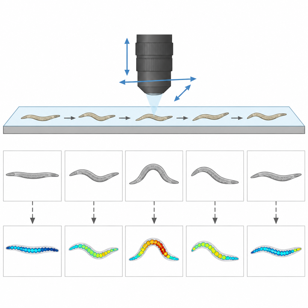
Following food cues and fine-scale nerve signals
Wormspy also handled more challenging scenes, such as worms crawling on a patch of bacteria that serves as food. On this bumpy, visually cluttered surface, the system still tracked a smell-sensing neuron called AWCON, revealing that its activity increases when a worm’s nose leaves the food, in line with theories about how animals search when food becomes scarce. Finally, the researchers pushed the resolution further by measuring tiny, localized calcium bursts in different segments of a single interneuron’s axon while the worm swung its head from side to side. They found that these signals were tightly linked to the direction and speed of head bends, and differed in timing from measurements taken in immobilized worms, highlighting the value of studying truly free movement.
Lowering the barriers to watching brains in motion
Taken together, these demonstrations show that Wormspy can connect detailed cell activity to natural behavior without expensive commercial microscopes or complex custom analysis. Because the design is modular, open, and built from standard parts, other labs can adapt it for different fluorescent markers, light-based stimulation methods, or even other small animals such as fly larvae. For non-specialists, the key message is that tools like Wormspy make it easier for researchers around the world to watch living nervous systems at work while animals behave as they normally would, bringing us closer to understanding how patterns of activity inside tiny brains create the rich actions we see on the outside.
Citation: Wittekindt, S.N., Owens, H., Guisnet, A. et al. An epifluorescence microscope design for naturalistic behavior and cellular activity in freely moving Caenorhabditis elegans. Nat Commun 17, 4411 (2026). https://doi.org/10.1038/s41467-026-72709-w
Keywords: Caenorhabditis elegans, calcium imaging, open-source microscope, neural activity, freely moving behavior