Clear Sky Science · en
Using rotational integration of oblique interferometric scattering to track axial spatiotemporal responses of tubular membrane protrusions
Watching tiny cell bridges in action
Our cells constantly reach out with tiny tube-like extensions to touch, sense and talk to their surroundings. These delicate structures help immune cells swallow bacteria, cancer cells spread, and tissues grow and repair. Yet they are so small and fast-moving that even advanced microscopes struggle to follow them in three dimensions without harming the cells. This study introduces a new way of looking at these “cell bridges” in real time, without dyes, to better understand how living cells connect and communicate.
Why current microscopes miss the full picture
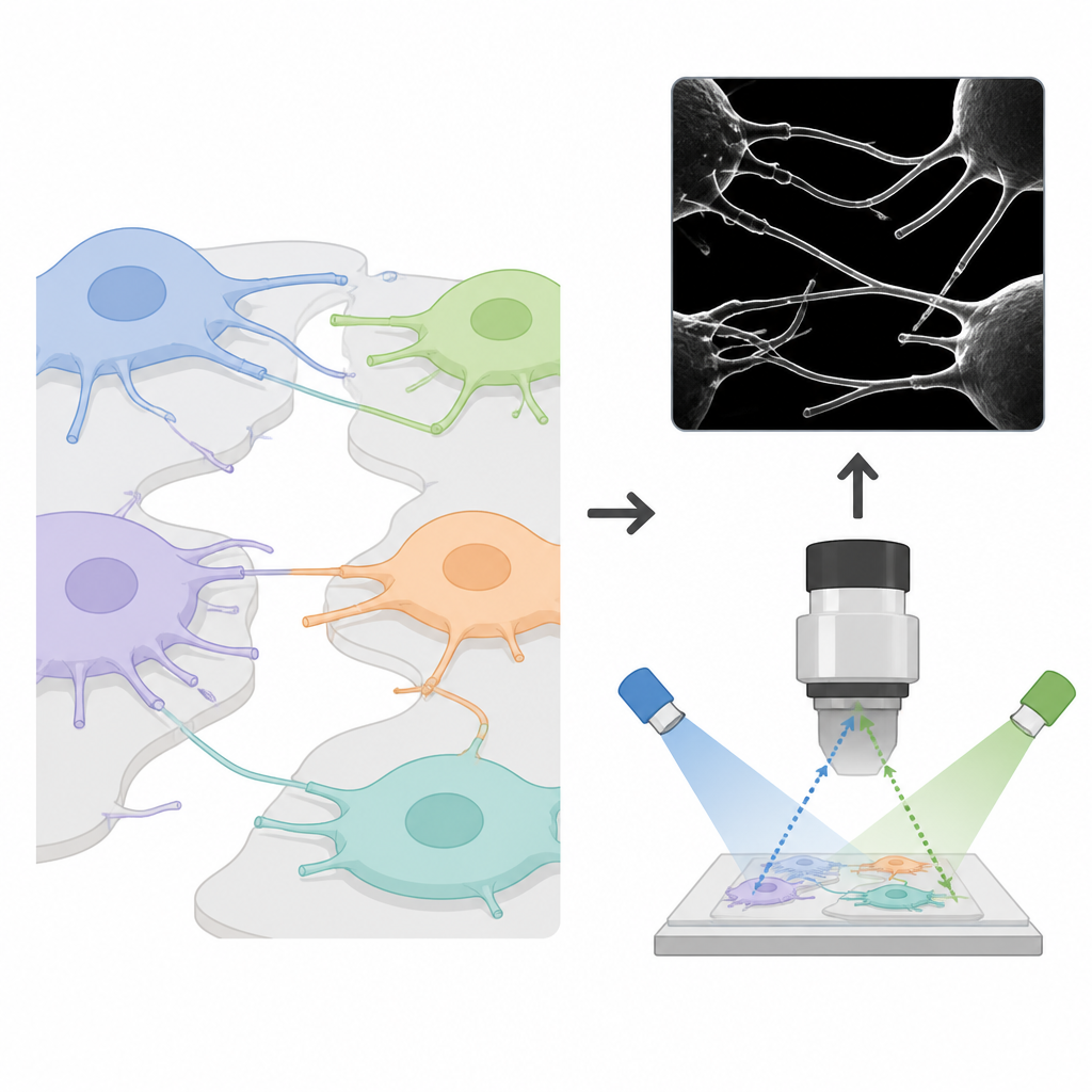
Many well-known cell protrusions, such as filopodia, tethers, trails and bridges, share similar shapes when seen from above but carry very different jobs. Some cling tightly to the surface a cell sits on, some poke upward to sense the environment, and others form long suspended tubes that can pass cargo between distant cells. Conventional light microscopes, especially those that rely on glowing fluorescent tags, have poor sharpness in the vertical direction and can damage cells over time with intense light. Electron microscopes can reveal fine detail, but the sample preparation often distorts these fragile tubes, and only dead, fixed cells can be examined. As a result, scientists have lacked a simple way to watch these structures form, twist and disappear in three dimensions inside living, crowded samples.
A new twist on scattering light
The team built on a technique known as interferometric scattering microscopy, which detects light scattered by tiny objects as it interferes with light reflected from a glass surface. This approach is extremely sensitive to small vertical shifts, making it promising for tracking nanoscale movements. However, in practice it suffers from speckle, a grainy background created by out-of-focus scatterers, and usually needs careful background subtraction using separate reference images. The authors discovered that by shining the laser into the sample at an angle and rapidly rotating that angle in a circle, then letting the camera integrate over one full rotation, the speckle patterns from distant planes shift laterally and cancel out, while the signal from the in-focus plane remains sharp. They call this rotational oblique interferometric scattering, or RO-iSCAT.
Clearer, gentler views of living cells
Using a combination of computer models and experiments with nanoparticles, extracellular vesicles and several cell types, the researchers showed that RO-iSCAT can boost the signal-to-noise ratio of the interference pattern by about tenfold without any numerical background subtraction. It resolves metal particles only a few hundred nanometres apart and captures crisp interference rings whose brightness changes smoothly with vertical position, allowing depth to be calibrated. When applied to living cells, the method tracks the three-dimensional journey of a single vesicle as it diffuses near the surface, slows while being taken up, and then moves within the cell interior. Crucially, because RO-iSCAT is label-free and uses modest light levels, long recordings of more than 20 hours left unlabeled cells healthy and dividing, while fluorescently labeled cells in the same light showed signs of damage, underscoring that phototoxicity originates mainly from the dyes, not the illumination.
Decoding different kinds of cell protrusions
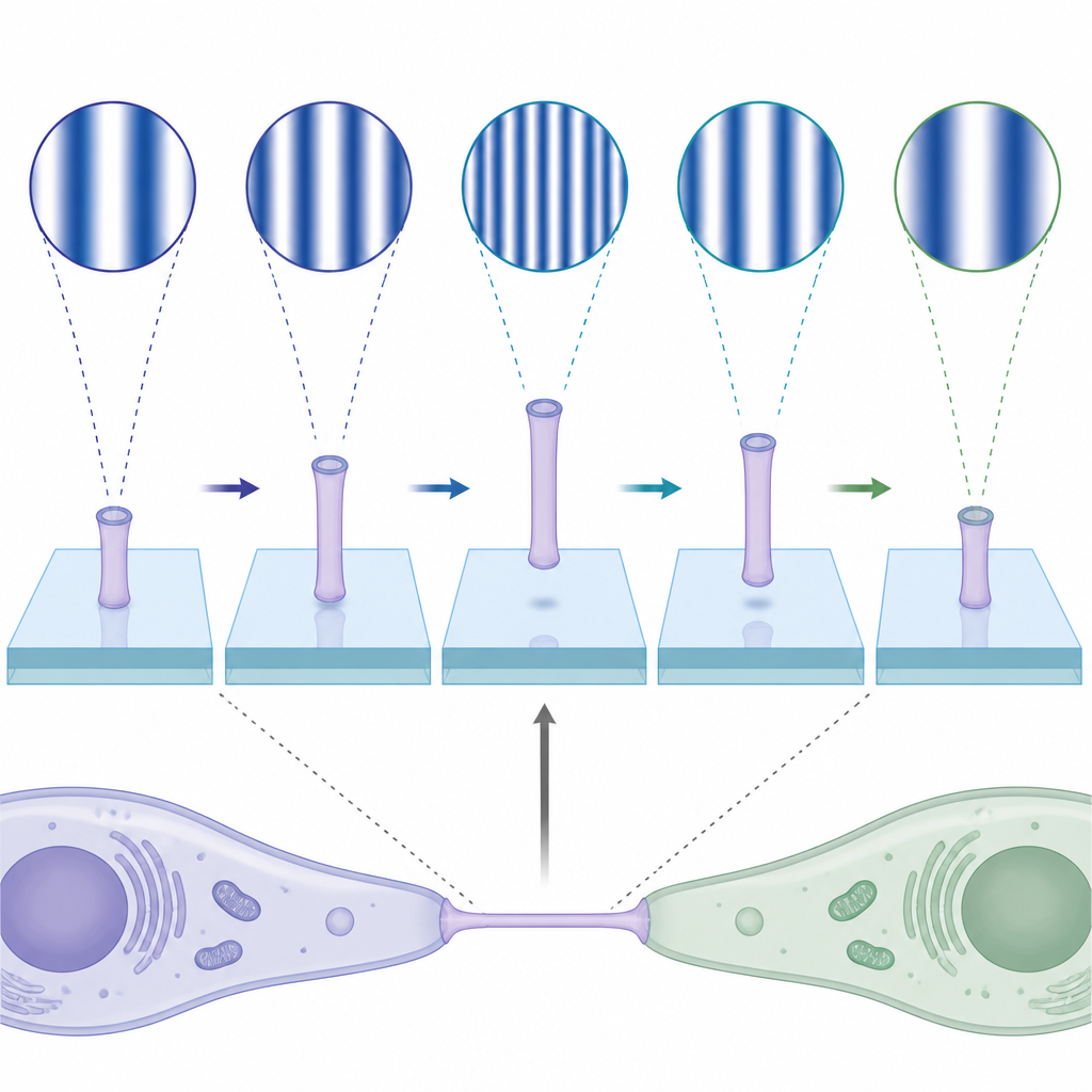
RO-iSCAT not only sees where slender membrane tubes are, it also captures how they move in the vertical direction over time. The team examined fibroblast and other cell types that naturally form a network of protrusions. They found that each kind of structure produces a distinct interference pattern: flat “trails” stuck to the glass show almost uniform contrast, downward-slanting “tethers” display closely spaced bright and dark bands, and suspended “bridges” between cells exhibit bands spaced farther apart along their length. By converting changes in brightness into vertical motion maps, they quantified that bridges show larger vertical fluctuations, like taut ropes vibrating in space, whereas trails and tethers move less. Counting these patterns across six cell types revealed that some cells, such as fibroblasts and connective tissue cells, make many more protrusions than, for example, insulin-secreting beta cells.
Following how cells connect with each other
The authors then turned to mixed cultures where fibroblasts communicate with each other or with pancreatic cancer cells. In one setup, they co-cultured fluorescently tagged and untagged fibroblasts and compared standard fluorescence imaging to RO-iSCAT. While fluorescence showed the overall outline of protrusions, RO-iSCAT exposed much richer behavior within the same tubes: neighboring protrusions that appeared static in fluorescence were seen to detach, twist and reattach, with their interference stripes changing rhythmically as they moved up and down. In a longer experiment, fibroblasts and cancer cells were seeded at opposite sides of a dish and allowed to grow toward each other. RO-iSCAT revealed how initially separate protrusions from each cell gradually merged into a single bridge, accompanied by a marked increase in vertical motion along the connection, a signature of mechanical strain that standard two-dimensional imaging could not capture.
What this means for understanding living tissues
In simple terms, this work provides a way to watch invisible threads between cells as they grow, tug and rearrange themselves in three dimensions, without adding dyes or harming the cells. By turning angled light and rotational averaging into a cleaner scattering signal, RO-iSCAT makes it possible to classify different kinds of cell protrusions by their optical fingerprints and to measure how lively or tense they are over time. This opens a path to studying how immune cells grab targets, how fibroblasts and cancer cells exchange signals, and how tissue networks form, all by reading subtle patterns in scattered light.
Citation: Liu, J., Lim, Y.J., Herrmann, D. et al. Using rotational integration of oblique interferometric scattering to track axial spatiotemporal responses of tubular membrane protrusions. Nat Commun 17, 4064 (2026). https://doi.org/10.1038/s41467-026-72302-1
Keywords: cell membrane protrusions, interferometric scattering microscopy, label free imaging, cell cell communication, 3D live cell imaging