Clear Sky Science · en
Eye movement kinematics reveal novel circadian organization of sleep substates
Why tiny fish eyes matter for our sleep
Sleep may feel like an all-or-nothing shutdown, but inside the brain it unfolds through a series of hidden stages. In mammals, some of these stages are defined by the way our eyes dart beneath closed lids. Until now, scientists were unsure whether such rich sleep structure existed in non-mammals like fish. This study uses high‑resolution recordings of larval zebrafish to reveal that their eyes also tell a surprisingly intricate story about sleep, one that is organized around the day–night cycle and echoes aspects of human sleep.
Watching fish around the clock
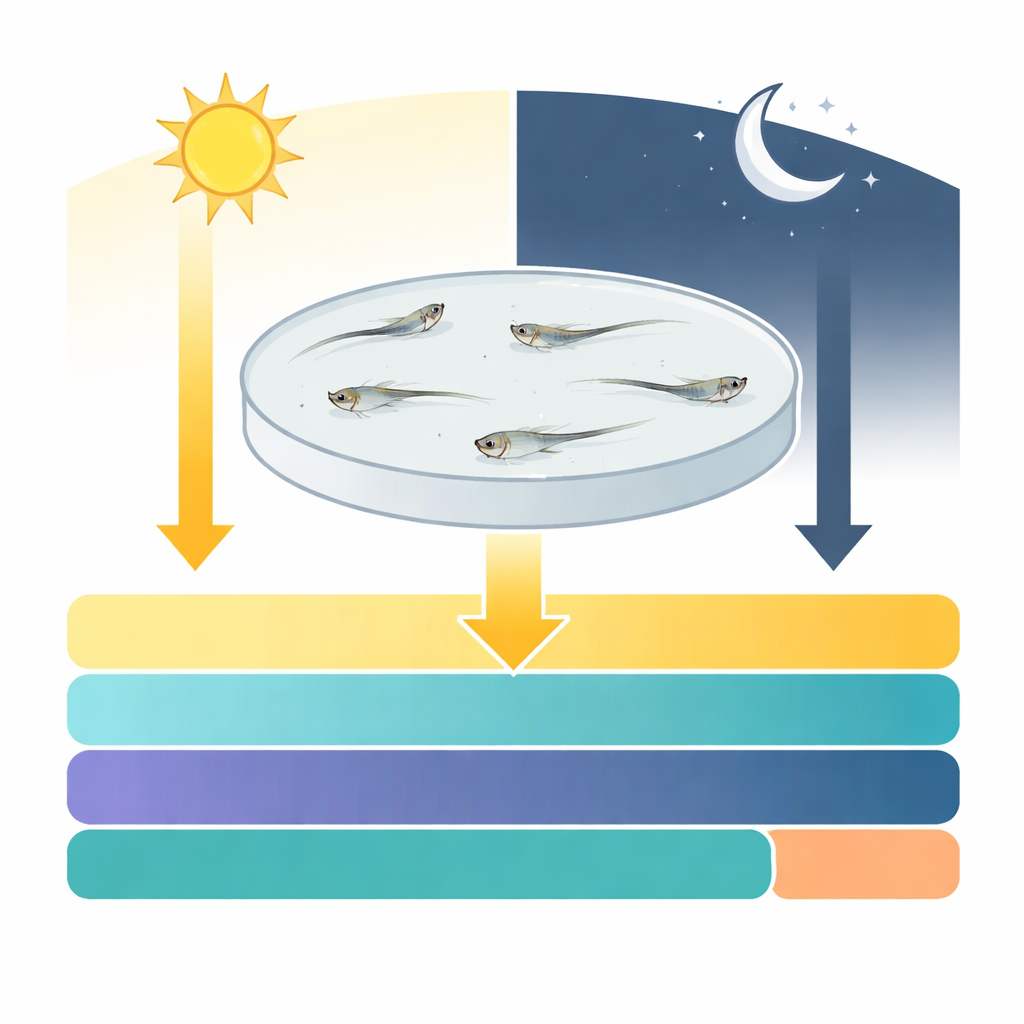
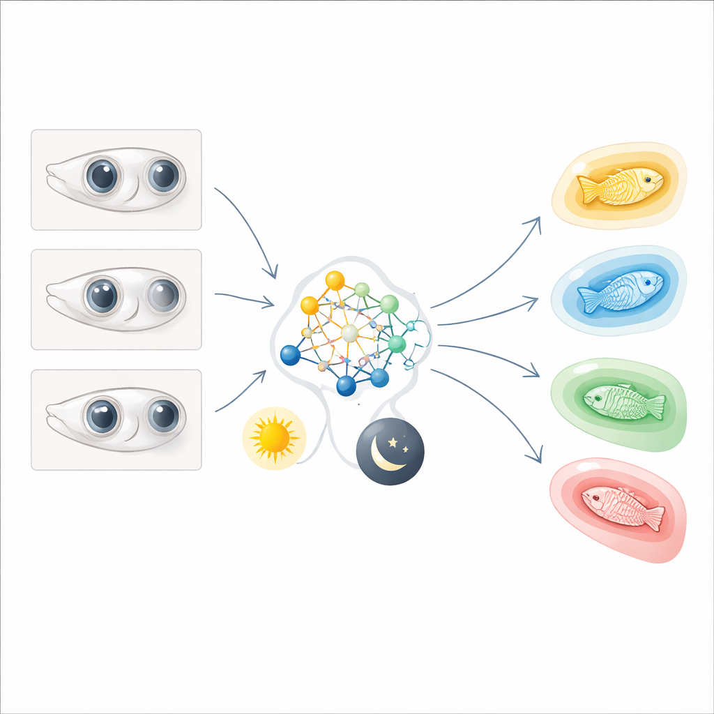
The researchers built an imaging system that can track up to 20 tiny zebrafish larvae at once, continuously, for days. The setup follows each freely swimming fish in a shallow circular dish, measuring both how fast it swims and how its eyes move. Periods when a fish barely moves for at least a minute count as “sleep” by established zebrafish criteria, because the animals are harder to wake. Within those quiet periods, the team analyzed how often the eyes made quick, coordinated jumps, how long they paused between jumps, and how smoothly they settled. This allowed them to sort every minute of sleep into distinct categories based purely on eye behavior.
Four flavors of fish sleep
From this massive dataset, four clearly separated sleep substates emerged. Three of them involved eye movements (called QEM-1, QEM-2, and QEM-3) and one showed no eye movement at all (QNEM). QEM-1 featured frequent, regular eye jumps with short, steady pauses, while QEM-2 and QEM-3 showed rarer and more irregular movements with different patterns of slowing and fixation. Crucially, all four substates were true sleep: in each, the fish were less likely to startle in response to bright flashes or mechanical taps than when they were awake. One substate, QEM-1, also showed partial loss of body posture, rebound after being deprived, and overall reduced activity in a key alertness center of the brain, confirming it as a genuine, low‑arousal sleep mode.
Sleep that follows the sun and the light
The four substates were not scattered randomly through the day. Instead, they followed strikingly different schedules tied to both the internal body clock and the surrounding light. QNEM and QEM-3 dominated during the night, providing deep, quiet sleep. QEM-2 was mostly a night state as well but became more common toward morning, suggesting a bridge toward wakefulness. Surprisingly, QEM-1 appeared almost only during the day and made up the bulk of daytime sleep. When the team kept the fish in constant light or constant darkness, the overall amount of sleep shifted, but the relative timing of these substates still showed clear circadian patterns. A simple artificial neural network model, fed only with time of day, light level, and how long the fish had been in the chamber, could reproduce most of the observed substate patterns, implying that a few key signals are enough to steer the system.
Shared sleep architecture across fish and inside the brain
These newly identified sleep substates were not a quirk of one laboratory strain. Closely related Danio species and multiple zebrafish strains all showed the same four substates and broadly similar day–night organization, with some species-specific twists. Zooming in on the brain, the authors used whole‑brain calcium imaging to watch neuronal activity during QEM-1. Most of the brain quieted down, including the noradrenergic locus coeruleus, a hub of wakefulness. Yet a small set of neurons in specific brainstem regions ramped their activity up or down in reliable patterns over the course of each QEM-1 episode. When the authors analyzed this activity across many neurons at once, they found that the brain’s trajectory during QEM-1 followed a smooth, low‑dimensional path, so consistent that a simple decoder could estimate how far along a QEM-1 bout had progressed using neural signals alone.
What this means for understanding sleep
To a lay observer, a resting zebrafish larva may seem simply asleep or awake. This work reveals that, beneath the surface, its sleep is broken into multiple, conserved stages distinguished by eye movements and tightly scheduled by circadian time and light. One daytime substate, QEM-1, shows all the hallmarks of sleep—from high arousal thresholds to homeostatic rebound and organized brain dynamics—despite occurring under bright light when we usually expect animals to be active. Together, these results suggest that richly structured, multi‑stage sleep is not unique to mammals. Instead, it may be an ancient feature of vertebrate brains, built from compact circuits that coordinate eye movements, posture, sensory responsiveness, and internal timekeeping.
Citation: Choudhary, V., Heller, C.R., Aimon, S. et al. Eye movement kinematics reveal novel circadian organization of sleep substates. Nat Commun 17, 4068 (2026). https://doi.org/10.1038/s41467-026-72222-0
Keywords: zebrafish sleep, circadian rhythms, eye movements, brain states, neural dynamics