Clear Sky Science · en
UltraFast Layer-Resolved Encoding (uFLARE) functional MRI deciphers bidirectional signaling from spontaneous activity
Listening In on the Brain’s Hidden Conversations
Even when we sit quietly, our brains buzz with internal chatter. Scientists know that this ongoing activity shapes how we see, feel, and recover from injury, but they have struggled to tell which signals move "up" from incoming sensations and which move "down" from higher brain areas that add expectations and context. This study introduces a new brain-scanning approach that can, for the first time, noninvasively tease apart these two directions of information flow, offering a window into how healthy brains work and how they reorganize after damage.
Two Roads Through the Thinking Brain
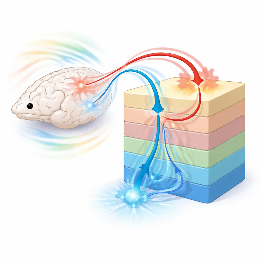
Our senses talk to the brain through a layered sheet of tissue called the cortex. Signals traveling from the eyes, ears, or skin into the brain are often called bottom-up: they carry raw data from the outside world into early processing areas and then up to more complex hubs. Top-down signals run the opposite way. They carry predictions, attention, and prior knowledge from higher areas back to early ones, tuning what we perceive. Until now, scientists needed invasive tools, such as electrodes inserted into the brain, to tell these two directions apart with fine detail. Conventional, noninvasive scans like standard fMRI can show where activity is happening, but not which way information is flowing within the thin stack of layers that make up the cortex.
A New Way to Read the Layers
The authors developed UltraFast Layer-Resolved Encoding, or uFLARE, a method that combines very rapid fMRI data with a mathematical model of how one brain region pools signals from another. Instead of simply tracking how strongly two areas are connected, their “layer-based connective field” model estimates how widely each point in the cortex draws information from its partners and how this pooling changes from the surface down to deeper layers. Because different layers are known from anatomy to specialize in either incoming or feedback signals, the pattern of pooling across depth can reveal whether a connection is mainly bottom-up or top-down. Using ultra-high-field MRI in rats, the team achieved both fine spatial detail across layers and fast sampling in time, allowing them to capture subtle, spontaneous fluctuations that carry directional information even in the absence of any external stimulus.
Distinct Fingerprints for Upward and Downward Signals
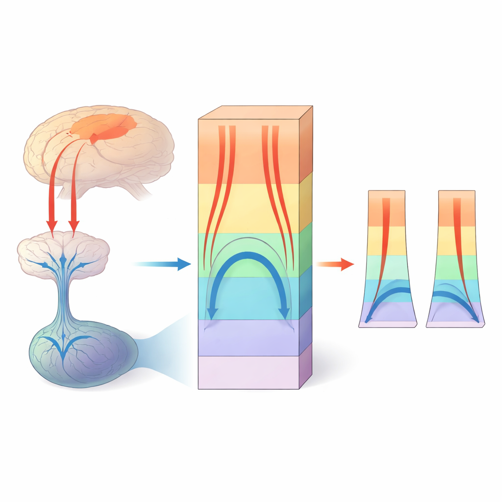
When the researchers examined how deep structures feeding the visual cortex connect into the layered sheet, they found a striking pattern. Connections that carried sensory input into “first-stop” visual areas showed the largest pooling in the middle layer, forming an inverted U-shaped profile across depth. In contrast, feedback connections from higher visual areas favored the top and bottom layers, tracing a U-shaped profile. These distinct shapes appeared not only during visual stimulation but also during spontaneous activity, indicating that bottom-up pathways are active even in the dark and that the brain’s internal conversations constantly rehearse both incoming and predictive signals. Similar layer-specific profiles emerged in touch and movement regions, suggesting a general organizing rule across sensory and motor systems.
Watching Circuits Adapt After Injury
The team then asked whether uFLARE could reveal how the brain rewires after damage. They created targeted lesions in primary visual cortex, mimicking a form of cortical blindness, and scanned the affected animals. As expected, normal input from the eye’s relay station (the lateral geniculate nucleus) into the destroyed visual area nearly vanished. But a new inverted U-shaped profile emerged from that same relay into higher visual regions, indicating that signals were now bypassing the damaged area and reaching downstream regions directly. A separate pathway that normally relays through another thalamic hub also changed its layer pattern, consistent with a broader reshaping of visual circuits. These observations align with prior invasive studies and with human “blindsight,” where people with primary visual cortex damage can still respond to visual cues they do not consciously see.
Why This Matters for Brains and Health
By showing that fast, layer-sensitive fMRI can distinguish bottom-up and top-down traffic from spontaneous activity alone, uFLARE opens a path to mapping the brain’s internal dialogue across entire networks without surgery or implanted devices. In the future, similar strategies at high-field clinical scanners could help doctors probe how perception, attention, and prediction go awry in conditions such as schizophrenia, autism, depression, or after stroke. Being able to noninvasively track how upward sensory evidence and downward expectations balance each other—and how that balance shifts as circuits adapt—could guide new therapies aimed at restoring healthy communication in the brain.
Citation: Carvalho, J., Fernandes, F.F., Valente, M. et al. UltraFast Layer-Resolved Encoding (uFLARE) functional MRI deciphers bidirectional signaling from spontaneous activity. Nat Commun 17, 3823 (2026). https://doi.org/10.1038/s41467-026-71506-9
Keywords: bottom-up and top-down signaling, layer-specific fMRI, spontaneous brain activity, cortical plasticity, connectivity modeling