Clear Sky Science · en
Manifold topological deep learning for biomedical data
Seeing Patterns Beneath Medical Images
Modern hospitals collect huge numbers of scans, from X rays to 3D MRIs, and we rely on computers to help doctors read them. This study introduces a new way for artificial intelligence to look at medical images that pays attention not just to pixel values, but also to the hidden shapes and flows inside each image. The goal is to make computer diagnoses more accurate, reliable, and easier to understand.
From Pictures to Smooth Surfaces
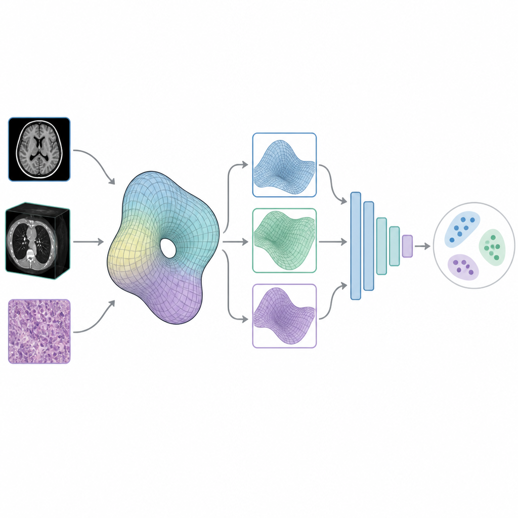
Most image analysis systems treat a scan as a flat grid of colored dots. The authors argue that many medical images are better viewed as smooth surfaces that bend and curve in space, much like the surface of a drum or the skin of a balloon. In mathematics, these smooth shapes are called manifolds. By treating an image as a manifold instead of a simple grid, the model can capture information about how structures are connected, where they loop around, and how different regions relate to each other in a continuous way.
Letting Flows Reveal Hidden Structure
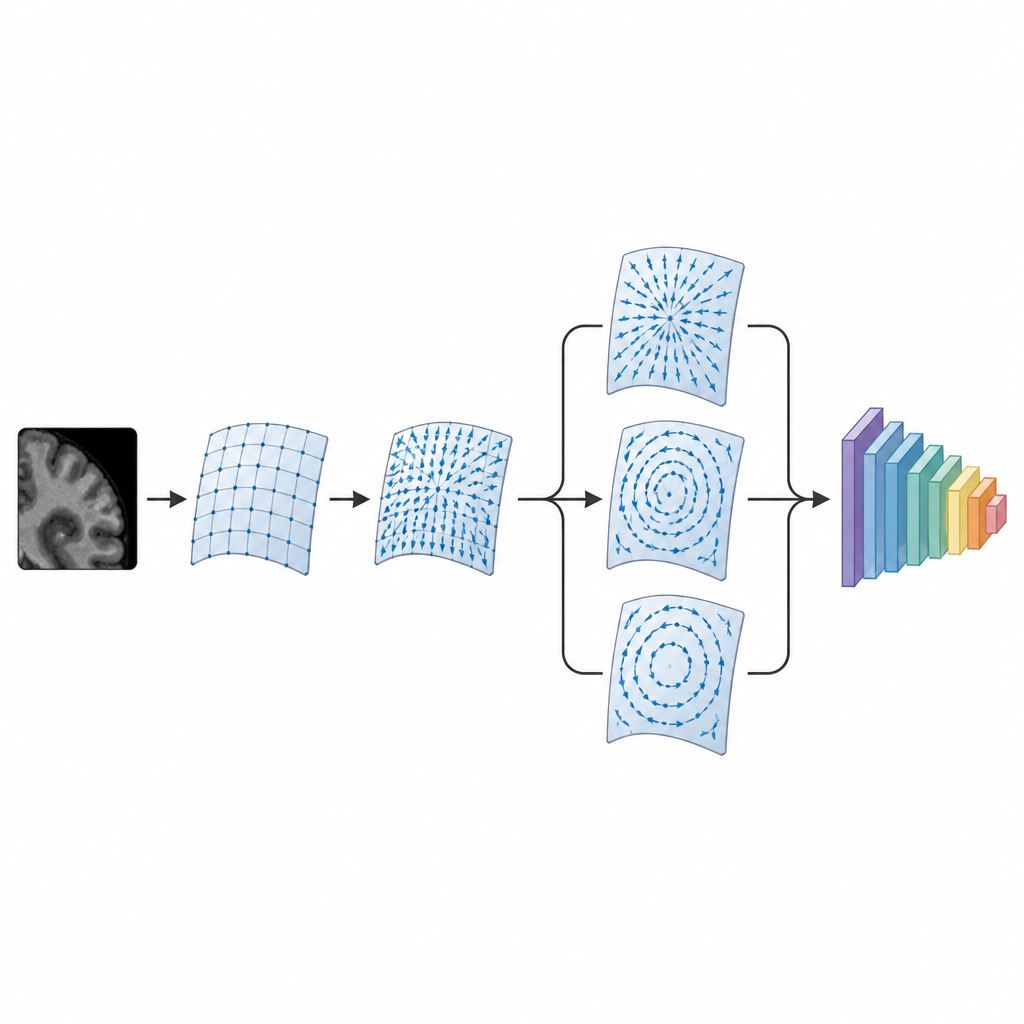
To dig into this manifold view, the method first builds a kind of flow field on top of each image, where tiny arrows describe how intensity changes from place to place. Using a branch of mathematics called Hodge theory, this flow is then split into three clean parts that do not interfere with one another. One part captures sources and sinks, another captures swirling patterns, and a third reflects large scale, global structure. This triple view turns a raw scan into a richer set of layers that emphasize different aspects of anatomy and tissue patterns.
Feeding Smarter Inputs to a Compact Network
After this mathematical reshaping, the three flow components are stacked together and passed into a small, carefully designed convolutional neural network. Unlike many popular medical AI models that contain tens of millions of adjustable weights, this network uses well under a million. Despite its compact size, it benefits from being fed input that is already organized according to shape and connectivity, instead of making the network discover these patterns from scratch. The authors tested their system on MedMNIST v2, a large benchmark of more than 700,000 biomedical images that span 2D and 3D scans and a wide range of organs, imaging devices, and task types.
Stronger Results Across Many Kinds of Data
The new approach outperformed leading deep learning models on almost all of the 17 MedMNIST datasets, including both 2D slices and 3D volumes. It did especially well on skin images, eye scans, blood cell slides, and 3D organ and neuron data, often achieving noticeably higher accuracy and better ranking of diseased versus healthy cases. The model remained strong when image sizes changed, when the number of training examples was small or large, and when the number of diagnosis categories varied. Tests on a real skin lesion collection at different resolutions showed that performance improved as more detail was available, but even low resolution images were handled better than by competing methods.
Why the Decomposition Matters
To check whether the mathematical splitting really helps, the authors repeated their experiments with a nearly identical network that skipped the manifold and flow decomposition and instead used the original images directly. In every case, the version with the decomposition performed better, sometimes by a wide margin. This suggests that the three flow based views capture complementary information about local texture and global shape that ordinary pixels alone do not provide, and that this extra structure makes it easier for the network to learn stable patterns.
A New Way to Read Medical Images
In plain terms, this work shows that teaching AI to respect the underlying shapes and flows in medical images can lead to more accurate and efficient tools for image based diagnosis. By combining ideas from geometry and topology with modern neural networks, the authors provide a framework that makes better use of the information already present in scans, while keeping the model relatively small. This manifold topological deep learning approach could help future systems read complex biomedical images more reliably, even when data are varied, limited, or noisy.
Citation: Liu, X., Su, Z., Shi, Y. et al. Manifold topological deep learning for biomedical data. Nat Commun 17, 4710 (2026). https://doi.org/10.1038/s41467-026-71392-1
Keywords: medical imaging AI, deep learning, topological data analysis, image classification, biomedical data