Clear Sky Science · en
Architecture and regulation of nanoscale chromatin domains
How DNA folds into tiny control hubs
Inside every human cell, two meters of DNA must fit into a nucleus smaller than a speck of dust, yet still stay readable so the cell can turn genes on and off. This article explains how DNA packs into tiny clumps, called nanoscale chromatin domains, that act like miniature control hubs. Understanding these hubs helps explain how cells develop, respond to stress, and drift into disease such as cancer, and may point to new ways to diagnose and eventually target those changes.
Seeing DNA structure beyond the limits of light
For decades, scientists thought DNA wrapped around proteins and then folded into a regular 30-nanometer fiber, like a neatly coiled rope. That picture has now been overturned. New super-resolution microscopes can see far below the usual blur of light, revealing that chromatin, the mix of DNA and proteins, instead forms irregular chains that bunch into dense clusters just 50 to 200 nanometers across. These nanoscale chromatin domains have now been observed by several advanced light and electron imaging methods across many mammalian cell types, suggesting they are a basic building block of genome organization. 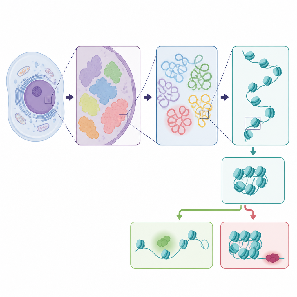
Inner core, active rim
Looking more closely, the authors describe how each tiny chromatin domain is organized like a layered sphere. The inner core tends to be tightly packed and enriched in chemical tags associated with gene silencing. The outer rim is looser, more accessible, and decorated with tags linked to active genes. Key proteins that turn genes on, including the cell’s main copying enzyme, cluster near these rims, at the boundaries between dense chromatin and the surrounding space. Other proteins, such as linker histone H1, help tighten the packing of these domains, while gaps between nucleosomes and certain chemical marks loosen them. Similar compact domains are also found at the nuclear edge, where chromatin sticks to the nuclear shell or wraps around the nucleolus, again following the pattern of dense, quiet cores and more active borders.
The hidden physics that shapes DNA clusters
The review then turns to how these tiny domains form and maintain their size. Physical models and computer simulations suggest they arise from a tug-of-war between simple diffusion, which makes similarly tagged pieces of chromatin clump together, and energy-consuming chemical reactions that add or remove those tags. Left alone, diffusion would make ever larger droplets, but enzymes that add activating marks or erase repressive ones continually chip away at growing clusters, freezing their size at the nanoscale. The cell’s transcription machinery adds another layer: as genes are read, mechanical forces and twisting of DNA can peel material off domain surfaces and change their size. When chromatin is tethered to the nuclear shell or nucleolus, additional surface forces help shape larger peripheral domains, much like droplets spreading on a surface in everyday liquids. 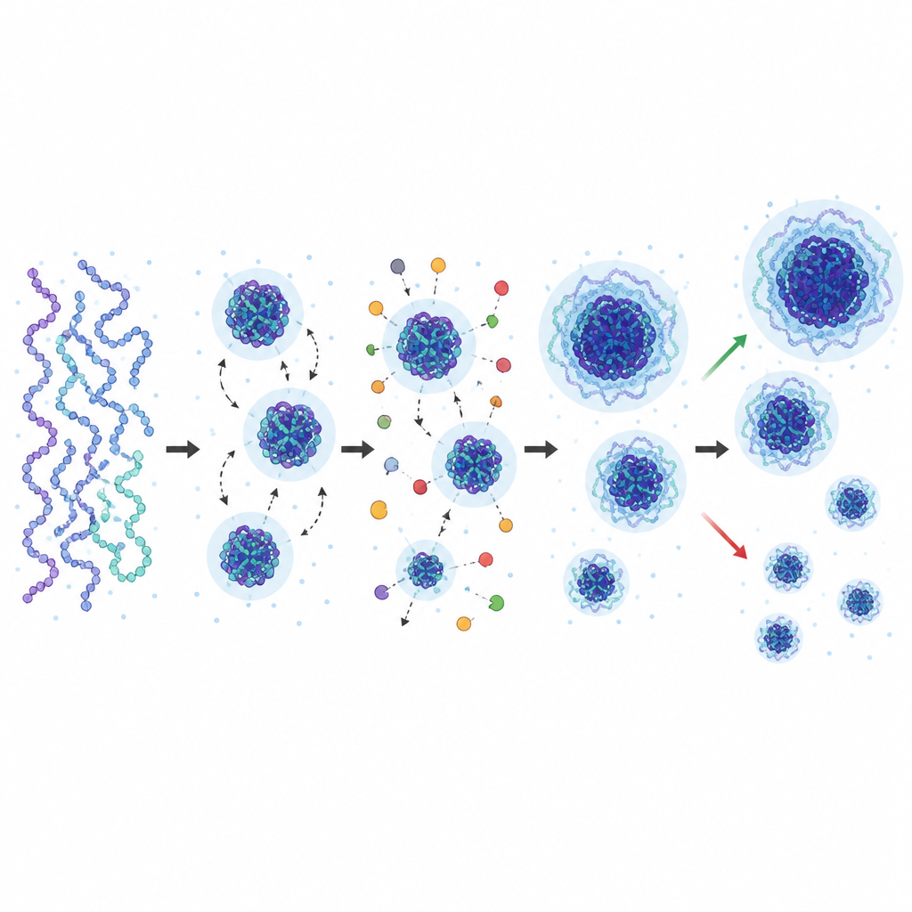
From stem cells to cancer: why domain size matters
Because all this activity is concentrated at domain boundaries, genes near the rims are especially sensitive to shifts in chemical reaction rates and mechanical forces. Studies show that soft or low-oxygen environments, aging, and tissue damage can enlarge domains and thicken peripheral layers, reducing access to many genes. During development, stem cells start with small, loose domains that favor flexibility, then consolidate into larger, denser domains as they commit to a fate. Reprogramming cells back to a stem-like state reverses this trend, with local domain loosening appearing before key genes switch on. In immune cells, activation breaks up domains and expands active space to support rapid bursts of gene expression. In many cancers, domains gradually lose their compact cores and fragment, increasing overall gene-accessibility and plasticity, a shift that seems to precede full-blown malignancy and drug resistance.
Why tiny domains hold big clues for health
Altogether, the article argues that nanoscale chromatin domains are not minor details, but central regulators that connect physical packing of DNA to gene activity and cell identity. Their size, position, and internal layering reflect a balance among chemical reactions, mechanical forces, and attachments to nuclear structures. When this balance shifts, cells can either become more flexible, as in stem cells, immune responses, and early cancer, or more locked in, as in mature tissues and some degenerative diseases. By viewing these tiny domains as tunable control units, the authors outline a path toward linking fine-scale DNA structure with development, aging, and disease, and suggest that these features could eventually help guide diagnosis and targeted intervention.
Citation: Vinayak, V., Lakadamyali, M. & Shenoy, V.B. Architecture and regulation of nanoscale chromatin domains. Nat Commun 17, 4682 (2026). https://doi.org/10.1038/s41467-026-71213-5
Keywords: chromatin domains, genome organization, epigenetics, super resolution microscopy, cell identity