Clear Sky Science · en
Structural basis for the carboxylation and epoxidation of human gamma-glutamyl carboxylase
How a vitamin helps blood and bones
Vitamin K is best known from the label on a multivitamin bottle, but inside our cells it powers a tiny machine that keeps blood clotting, bones hard, and arteries flexible. This machine, an enzyme called gamma glutamyl carboxylase (GGCX), quietly fine tunes many proteins before they are allowed to work. The study behind this article uses high resolution imaging to reveal how GGCX recognizes different protein partners and uses vitamin K to modify them, helping explain both healthy biology and certain bleeding and calcification disorders.
The body’s vitamin K workshop
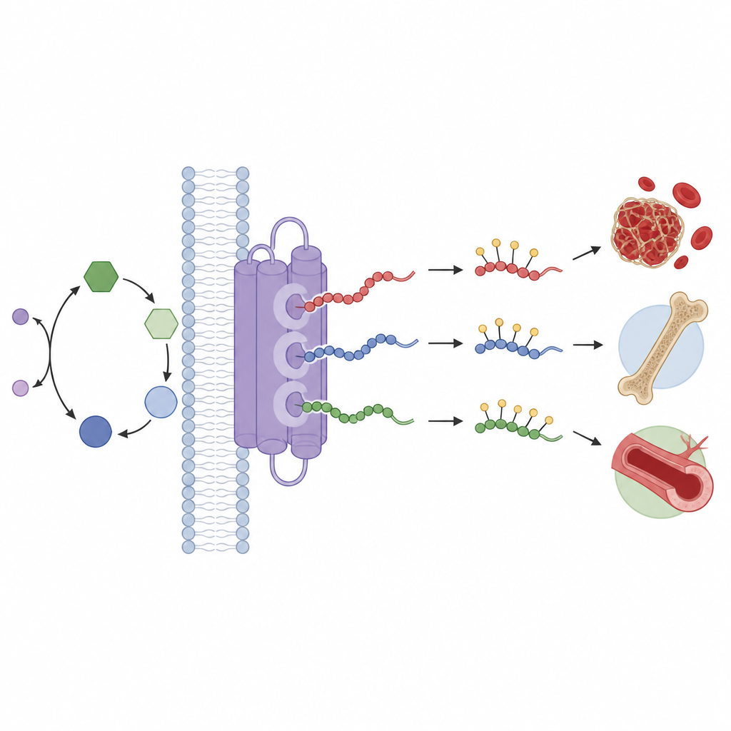
GGCX sits in the membrane of the cell’s protein processing factory, the endoplasmic reticulum. Its job is to add small chemical groups to specific building blocks, called glutamates, on a family of vitamin K dependent proteins. These targets include classic blood clotting factors as well as proteins that shape bone and prevent unwanted mineral deposits in blood vessels. Without this finishing step, clotting factors do not work properly and bleeding can occur, while other tissues may lose protection against calcification. The enzyme draws energy from the vitamin K cycle, in which vitamin K is repeatedly altered and recycled, and this cycle is also the target of the common drug warfarin.
Seeing the enzyme and its partners
To understand how GGCX does its job, the researchers used cryo electron microscopy, a technique that freezes proteins in thin ice and images them with electrons. They produced human GGCX together with five different natural partners: two clotting factors and three proteins not directly involved in coagulation. The resulting images reached near atomic detail, allowing the team to build 3D models of the enzyme docked with each partner. All complexes showed the same basic arrangement: a cluster of nine membrane spanning helices forming the core and a luminal head made of three subdomains that grabs the front “propeptide” segment of each client protein.
How GGCX picks out its clients
The study shows that the short propeptide acts as an ID tag that guides each protein to GGCX. Three neighboring patches on the enzyme form a recognition site that embraces a series of mostly hydrophobic amino acids at fixed positions along the propeptide. These key residues are strongly conserved among vitamin K dependent proteins, and delicate mutational tests confirmed their importance. When the team swapped these contact points for less favorable amino acids, the ability of the propeptide to boost vitamin K use and support the reaction dropped sharply. Small differences at one of these positions help explain why certain proteins, such as the bone hormone osteocalcin, bind more weakly to GGCX than clotting factors do.
Three ways into the reaction center
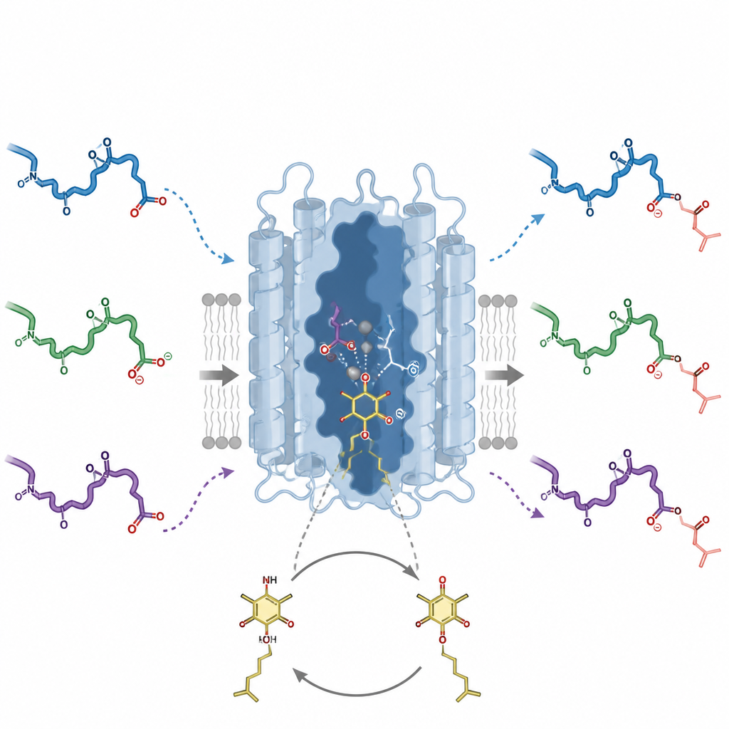
Although GGCX recognizes all propeptides in a similar way, the business end of each client protein, the glutamate that will be modified, can approach the active site by three distinct routes. In one mode, seen for clotting factors and a proline rich protein, the target glutamate lies just beyond the propeptide. In a second mode, used by osteocalcin, an extra segment on the far side of the reaction site also tethers the protein to the enzyme and supports efficient processing. A third, newly revealed mode occurs in matrix Gla protein, which helps prevent vessel calcification: here the reactive glutamate sits before the propeptide rather than after it. A flexible but length sensitive linker thread guides this unusual site into the same catalytic pocket; shortening it cripples the reaction even if the chemistry site is intact.
Coupling vitamin K use to protein repair
The high resolution structures also captured vitamin K in the form of its epoxide product nestled in the reaction cavity directly beneath a reactive glutamate side chain. This snapshot, backed by mutational experiments, outlines how specific amino acids in GGCX coordinate both vitamin K and the glutamate to choreograph two linked reactions: oxidation of vitamin K and attachment of carbon dioxide to the protein. Additional structures with only the propeptide present suggest how the enzyme shifts from an idle state to a fully engaged form as the full substrate binds. Together, these insights explain how one membrane embedded machine can read a family of related ID tags, steer chemically diverse protein segments into a shared pocket, and use vitamin K to tune factors that control clotting, bone quality, and vascular health.
What this means for health and disease
By revealing the detailed shapes and movements of GGCX and its partners, this work clarifies how vitamin K is harnessed to activate proteins throughout the body. It explains how different client proteins can be loaded in several ways into a single reaction center, and how subtle changes in docking points or linker length can weaken modification. These structural blueprints may help researchers interpret disease causing mutations in GGCX or its clients, refine our understanding of warfarin like drugs, and ultimately guide efforts to modulate vitamin K dependent processes in clotting, bone disease, and vascular calcification.
Citation: Zhang, W., Chen, Q., Zhang, B. et al. Structural basis for the carboxylation and epoxidation of human gamma-glutamyl carboxylase. Nat Commun 17, 4492 (2026). https://doi.org/10.1038/s41467-026-71212-6
Keywords: vitamin K, gamma glutamyl carboxylase, blood clotting, bone metabolism, vascular calcification