Clear Sky Science · en
Whole-proteome phage immunoprecipitation sequencing reveals germ cell tumor–specific immunosignature
Why this research matters to everyday health
Testicular germ cell tumors are the most common solid cancers in young men, yet the blood tests doctors rely on can miss many cases or give confusing results. This study introduces a new way to read the immune system’s fingerprint in the blood, with the goal of spotting these tumors more accurately and helping doctors choose the right treatment sooner.
Limits of today’s tumor blood tests
For decades, doctors have measured a handful of substances in the blood to help diagnose germ cell tumors and track treatment. These markers, such as beta HCG, AFP, and LDH, rise in only a portion of patients and can also be elevated for reasons unrelated to cancer. As a result, many men with normal test results may still harbor a tumor, while others may face uncertainty or overtreatment. Ultrasound scans of the testes are very good at finding suspicious lumps but cannot reliably distinguish tumor types or detect tiny spread elsewhere in the body.
Letting the immune system reveal hidden tumors
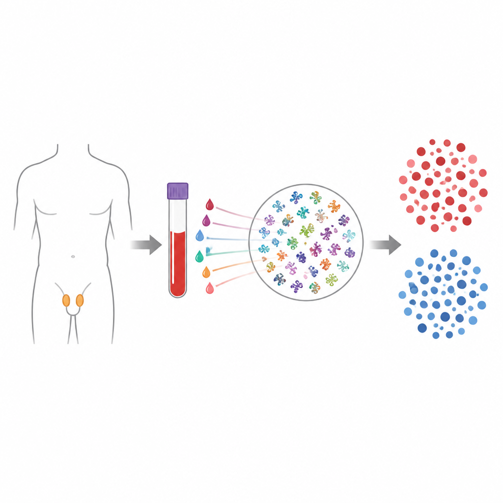
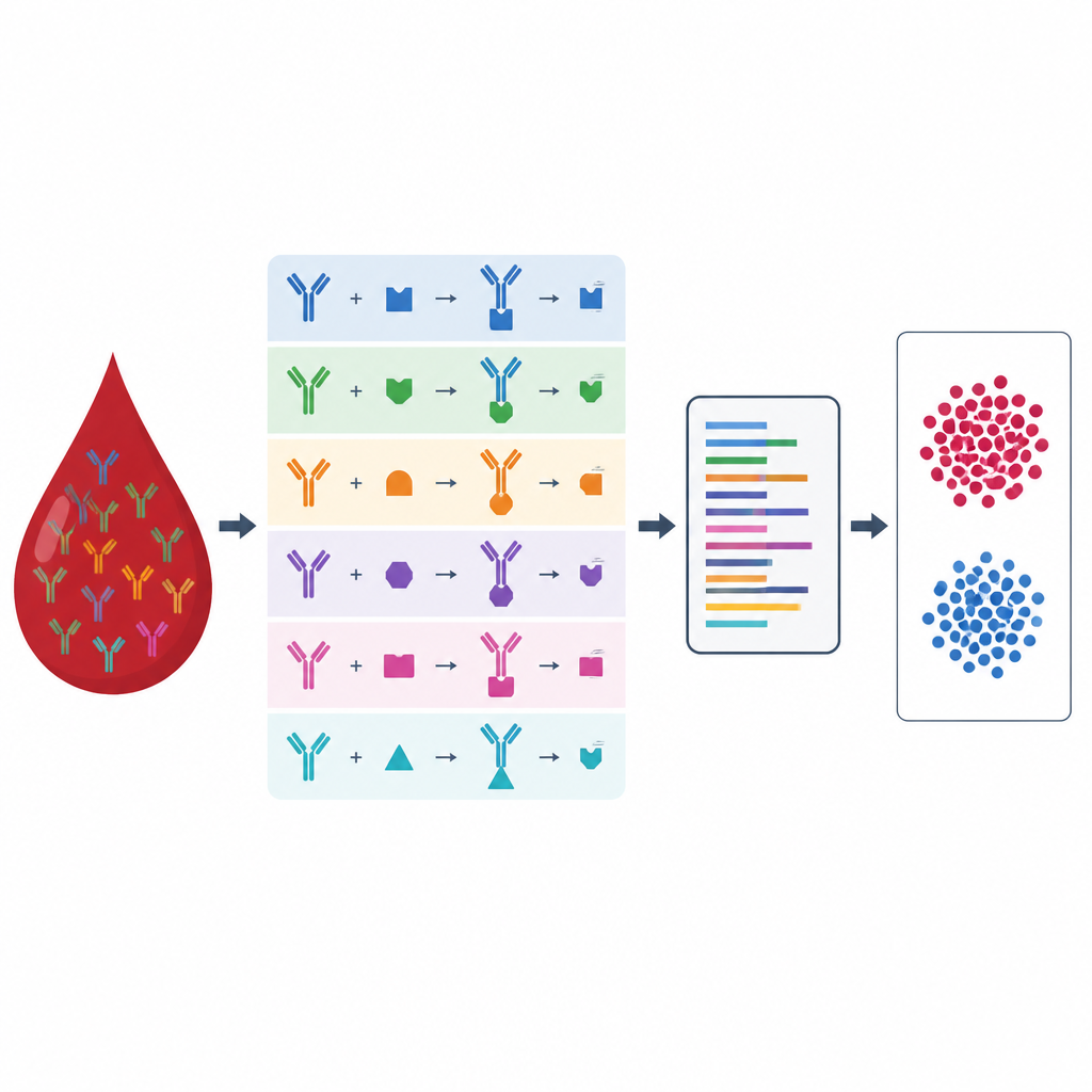
The researchers used a technology called phage immunoprecipitation sequencing, which can test blood against hundreds of thousands of tiny protein fragments that represent nearly all human proteins. When a person has a tumor, the immune system often produces antibodies that recognize unusual or overexpressed proteins in the cancer cells. By capturing and sequencing which protein fragments the antibodies latch onto, the team built an “immunosignature” that reflects the presence of a germ cell tumor. From 427 blood samples, including 150 patients with germ cell tumors and 277 controls, they trained and tested computer models to distinguish tumor cases from noncancer controls.
A new blood panel with a strong immune fingerprint
The main model, called GCT iSIGN, consists of 24 short protein fragments drawn from 16 different proteins. Many of these proteins belong to zinc finger families that help control gene activity, as well as viral-like elements and a surface molecule called MUC4. Together, this set of targets allowed the model to identify germ cell tumors with 93 percent sensitivity and 99 percent specificity, meaning it correctly detected most cancers while rarely flagging healthy or unrelated disease samples. Strikingly, it recognized 23 of 24 patients whose traditional markers were all normal, including many with early stage disease and pure teratoma, a subtype that current blood tests often miss.
Telling one tumor type from another
Because seminoma and nonseminoma tumors behave differently and are treated in different ways, the team also built a second model, Sem iSIGN, to distinguish these types using immune fingerprints alone. This panel uses 17 protein fragments from just five proteins, including LUZP4, a cancer testis antigen the group had previously linked to paraneoplastic disease. In validation testing, Sem iSIGN correctly identified seminoma with 96 percent specificity and 65 percent sensitivity. While not accurate enough to replace tissue examination under the microscope, it could complement pathology and standard markers in challenging cases.
Checking the signals in tumors and with standard lab tests
To ensure the immune fingerprints reflected real tumor biology, the scientists examined public RNA sequencing data from large cancer datasets. They found that many of the genes behind the GCT iSIGN and Sem iSIGN panels were expressed at much higher levels in testicular tumors than in kidney or prostate cancers, and that expression patterns differed between seminoma and embryonal carcinoma. They also used staining of tumor tissue and specialized antibody tests (ELISA) to confirm that several key targets, including ERVK7, MUC4, ZNF91, and LUZP4, are present in germ cell tumors and that blood antibodies detected by the new method align well with more traditional laboratory assays.
What this could mean for patients
This work shows that reading the immune system’s detailed response to cancer can provide a powerful, blood based signal of testicular germ cell tumors, even when standard markers appear normal. Although further prospective studies are needed before this approach becomes part of routine care, the immunosignature panels described here offer a scalable and relatively low cost path toward more accurate diagnosis, better tumor subtyping, and more tailored management for young men facing these cancers.
Citation: Hammami, M.B., Knight, A.M., Kherbek, H. et al. Whole-proteome phage immunoprecipitation sequencing reveals germ cell tumor–specific immunosignature. Nat Commun 17, 4733 (2026). https://doi.org/10.1038/s41467-026-71174-9
Keywords: germ cell tumor, testicular cancer, autoantibodies, cancer biomarkers, phage immunoprecipitation sequencing