Clear Sky Science · en
Energetic diversity in retinal ganglion cells is modulated by neuronal activity and correlates with resilience to degeneration
Why eye nerve cells and their energy use matter
The nerve cells that carry visual information from the eye to the brain work hard around the clock, burning large amounts of fuel to keep signals flowing. This study looks inside those cells in living mice to see how much chemical energy they hold, how they spend it during activity, and how these differences relate to whether the cells survive injury. The results show that not all retinal nerve cells manage energy in the same way, and that this hidden diversity may help explain why some cells are more resistant to damage than others. 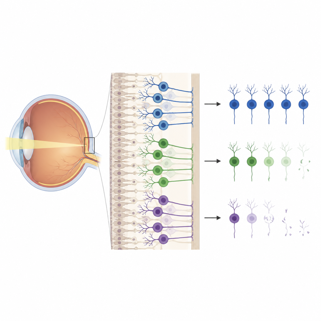
Different cells, different energy set points
The researchers focused on retinal ganglion cells, the output cells of the eye that send signals along the optic nerve. Although these cells share the same environment, they come in many types with distinct roles in visual processing. Using a fluorescent sensor delivered by a harmless virus, the team measured levels of ATP, the main energy currency of cells, in single ganglion cells in live mice. They found that ATP levels were not uniform: some cell types, including a fast-signaling group called alpha cells, sat at a lower steady energy level than others that respond to light in different ways. These differences were stable over time, showing that each cell type maintains its own preferred energy set point rather than all converging on the same level.
Testing how cells cope when fuel production is blocked
Next, the scientists challenged the cells by blocking key steps of mitochondrial energy production, the process that normally turns oxygen and nutrients into ATP. When they partially shut down parts of this machinery, ATP fell most sharply in alpha cells, especially in a subtype that is usually very active. Other ganglion cell types showed milder drops under the same conditions. Paradoxically, separate staining experiments showed that alpha cells actually have more mitochondria and higher amounts of energy-making proteins, suggesting they are built for high output and high demand rather than being weak. When the team blocked the final step of the energy chain, ATP fell more evenly across all cells, showing that some disruptions hit every cell while others expose the unique vulnerabilities of particular types. 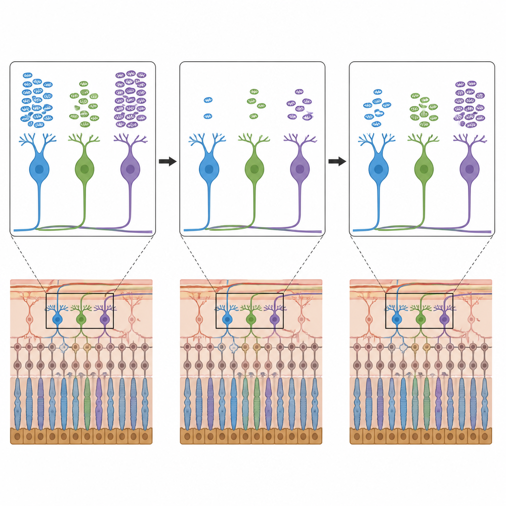
How nerve activity shapes energy use
The study also asked how day-to-day signaling affects energy balance. By using drugs to boost or dampen activity in the retinal circuit, the researchers could drive large, sustained changes in calcium signals, a readout of firing. Strong activation caused ATP to drop across the population at first, but levels then recovered toward baseline even while activity stayed high, implying that cells quickly ramp up energy production to match demand. Reducing activity led to a slow rise in ATP. Yet more natural, brief bursts of light-driven activity did not measurably change ATP, which indicates that under normal conditions the retina has enough energetic buffering and control to handle visual processing without draining its reserves. Importantly, when mitochondrial function was partially blocked, turning down activity protected ATP levels, and even pausing the imaging light briefly allowed some recovery before ATP fell again when stimulation resumed.
Energy levels and survival after optic nerve injury
To connect these patterns to disease, the team injured the optic nerve, a model for conditions such as glaucoma, and followed the same cells over two weeks. Surprisingly, the ganglion cells that survived long term had lower starting ATP levels than those that later died, both among alpha cells and among other types. After injury, many cells showed a temporary rise in ATP over several days before returning toward baseline, hinting at a stress response rather than simple energy failure. The researchers also tested daily treatment with a vitamin-like compound related to nicotinamide, which has been reported to help retinal cells in other models. This treatment boosted the post-injury ATP increase but did not significantly improve survival in this setting, suggesting that higher ATP on its own is not enough to guarantee protection.
What this means for protecting vision
Together, these findings reveal that retinal ganglion cells differ in how much energy they keep on hand, how they depend on mitochondrial fuel during activity, and how these traits relate to their chances of surviving damage. Cells with lower resting ATP are not necessarily weaker; in this study they were more resilient to injury, perhaps because they are adapted to handle energetic stress or limit harmful byproducts of energy production. Understanding this hidden metabolic diversity could guide future strategies to protect vulnerable vision pathways by tuning not just how much fuel cells receive, but how they manage and prioritize that energy over time.
Citation: Wang, Z., Zhao, C., Xu, S. et al. Energetic diversity in retinal ganglion cells is modulated by neuronal activity and correlates with resilience to degeneration. Nat Commun 17, 4531 (2026). https://doi.org/10.1038/s41467-026-71106-7
Keywords: retinal ganglion cells, neuronal metabolism, mitochondrial function, optic nerve injury, neurodegeneration