Clear Sky Science · en
Simultaneous profiling of native-state proteomes and transcriptomes of neural cell types using proximity labeling
Seeing Cells in Two Ways at Once
Every cell in the brain runs on instructions written in RNA and carried out by proteins, but these two layers do not always match. A gene can look quiet at the RNA level while its protein is abundant, or vice versa. This study introduces a method called SPARO that lets scientists read both the RNA and protein landscapes of specific brain cell types at the same time, and crucially, in their natural surroundings. For readers, this opens a window into how brain cells really behave in health, aging, and disease, beyond what can be seen from genes alone.
Why RNA and Protein Stories Do Not Always Match
For years, biologists have used RNA sequencing to see which genes are switched on, and mass spectrometry to catalog the proteins those genes produce. But many steps lie between an RNA message and a finished protein, including how long messages survive, how quickly they are translated, how stable proteins are, and where they are stored inside the cell. As a result, RNA and protein levels only modestly agree. Existing methods can look at both in simple cell cultures, but in living brain tissue they usually require breaking cells apart, sorting them physically, or focusing on a narrow slice of molecules, which can distort the very states scientists hope to measure.
A New Tagging Strategy Inside Living Cells
The team built SPARO around an engineered enzyme called TurboID, which acts like a molecular paint sprayer in the cell. When supplied with biotin, TurboID attaches tiny biotin tags to nearby proteins, many of which normally bind RNA or form part of the ribosome. Using a magnetic handle that recognizes biotin, the researchers can pull down these tagged proteins along with the RNA molecules attached to them. From the same starting material, they then split the haul: one part goes to protein analysis, the other to RNA sequencing. This creates a matched snapshot of which RNAs and proteins were present in the same cell type and at the same moment. 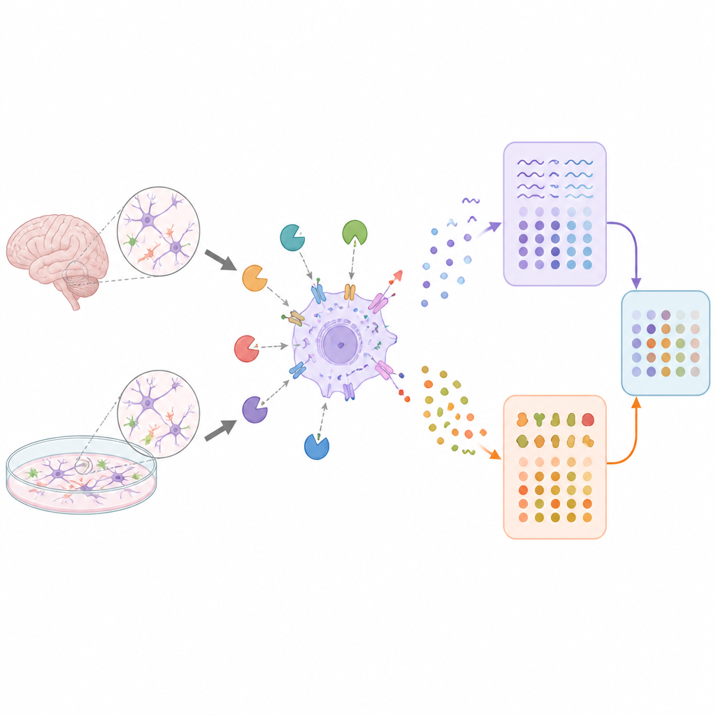
Testing the Method in Immune-Like Brain Cells
First, the researchers tried SPARO in a well-controlled dish model using BV2 microglial cells, which resemble the brain’s immune cells. They engineered these cells to express TurboID mainly in the cytosol, where much of the cell’s machinery resides. They then exposed the cells to a bacterial component that triggers inflammation and compared the RNAs and proteins captured by SPARO to traditional measurements from whole-cell extracts. The RNA profile from SPARO overlapped almost completely with the global RNA profile and accurately reported inflammatory gene responses. The protein profile was somewhat more selective, favoring cytosolic proteins and under-representing nuclear and mitochondrial components, but still recovered key inflammatory proteins and pathways.
Reading Neurons and Astrocytes in the Intact Brain
The real test was whether SPARO could work inside a living brain without isolating cells. The authors crossed mice carrying TurboID with mouse lines that turn on the enzyme only in either excitatory neurons or astrocytes, a major class of support cells. After giving the animals biotin in their drinking water, the team harvested the cortex and applied SPARO. The resulting protein and RNA profiles clearly separated neurons from astrocytes and were enriched for classic markers of each cell type. When they compared SPARO’s astrocyte RNA readout to that from RiboTag, a popular ribosome-based method, the two sets of transcripts strongly agreed, though SPARO captured a broader range of RNA-binding proteins and even small non-coding RNAs such as microRNAs. 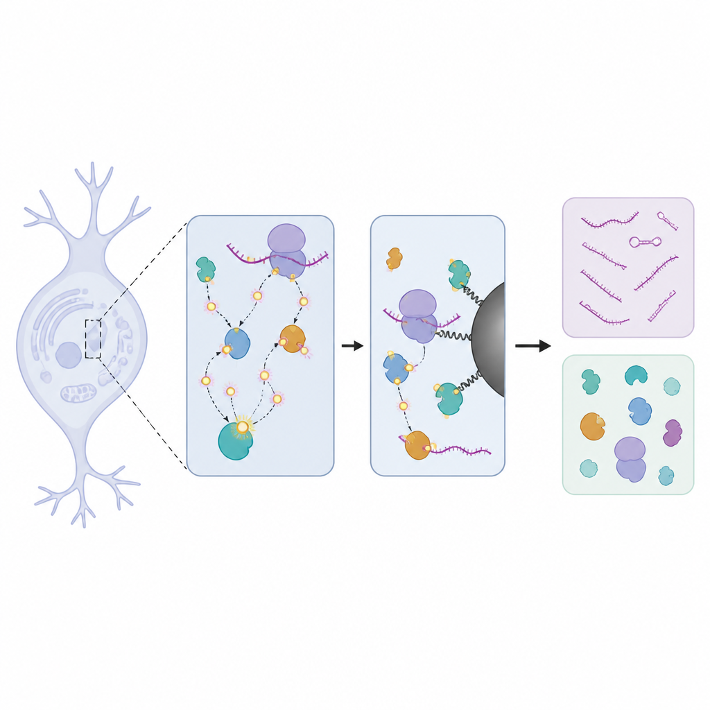
What Discord Between RNA and Protein Reveals
Having matched RNA and protein maps from the same cell types, the researchers asked where the two signals agreed and where they diverged. Across astrocytes and neurons, most genes fell into two camps: both RNA and protein were low, or both were high. But a sizeable minority were discordant. Genes with much more protein than RNA were often involved in the cell’s internal scaffolding, with astrocytes skewed toward microtubule components and neurons toward actin-related parts, reflecting their different shapes and roles. Genes with abundant RNA but relatively little protein were often linked to mitochondria and energy metabolism, hinting that these messages are transcribed but not fully translated or that their proteins are rapidly turned over. Similar patterns appeared in independent datasets and in human cell lines, suggesting that the mismatches are biological features, not artifacts of the tagging method.
Why This Matters for Understanding the Brain
To a non-specialist, the main message is that SPARO offers a way to listen to both the plans and the products inside specific brain cells without disturbing their native environment. The study shows that RNA measurements alone can miss important aspects of cell behavior and that systematic mismatches between RNA and protein levels follow meaningful patterns tied to cell type and cellular function. By making it possible to chart these relationships in neurons, astrocytes, and other cell types across the brain, SPARO sets the stage for richer maps of how cells change in development, aging, and neurological disease, and for choosing better RNA or protein markers when tracking those changes.
Citation: Ramelow, C.C., Dammer, E.B., Xiao, H. et al. Simultaneous profiling of native-state proteomes and transcriptomes of neural cell types using proximity labeling. Nat Commun 17, 4636 (2026). https://doi.org/10.1038/s41467-026-71098-4
Keywords: transcriptome, proteome, neural cell types, proximity labeling, TurboID