Clear Sky Science · en
Generative AI for misalignment-resistant virtual staining to accelerate histopathology workflows
Why smarter digital stains matter
When doctors diagnose cancer, they often rely on thin slices of tissue that have been soaked in colorful chemical dyes. These stains reveal the shapes and patterns that signal disease, but the process is slow, labor-intensive, and uses harsh reagents. This study explores how generative artificial intelligence can create realistic "virtual" stains on a computer instead, and shows how a new approach can avoid a long-standing technical roadblock that has held back this technology in real hospitals.
From glass slides to virtual color
In traditional pathology, different stains are used to highlight different features of tissue: one might show cell nuclei, another the supporting tissue, another specific proteins. Each stain requires extra time, chemicals, and sometimes additional pieces of precious patient tissue. Virtual staining takes a different route. It starts from an image that is easy to acquire, such as a label-free fluorescent picture or a common stain, and uses AI to predict how the same tissue would look with another stain. In principle, this could speed up diagnosis, save material, and reduce environmental impact. 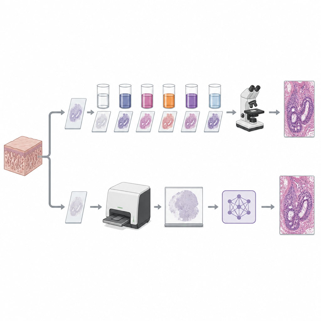
The hidden problem of tissue shift
In practice, virtual staining has struggled because real tissue moves and warps during chemical processing. Two images of what should be the same area rarely line up perfectly at the pixel level. Most AI methods need tightly matched pairs of "before" and "after" images to learn the right color transformation. If the tissue shifts even slightly, the models can blur structures, invent details, or lose important features. Some earlier techniques tried to work without matching pairs at all, while others added clever loss functions or built-in alignment steps, but they often traded structural accuracy for overall style, producing images that looked plausible yet were not trustworthy for diagnosis.
A two-part brain for alignment and color
The authors introduce a new framework called DGR, short for Decoupled Generation and Registration. Its key idea is to split the job into two cooperating AI modules: one focuses only on changing the appearance of the image to mimic the desired stain, while the other focuses only on aligning images that do not quite match in position. Extra constraints are added so that the virtual stain keeps the same shapes as the input image, while a separate alignment step quietly corrects for tissue shifts when comparing to real stained examples during training. By separating these roles, the system can learn from imperfectly matched data without being confused by it.
Putting the method to the test
The team tested DGR on five different datasets and four kinds of tasks: creating standard hematoxylin and eosin (H&E) stains from label-free fluorescence images, converting H&E into a more specialized kidney stain, generating advanced multiplex protein stains from routine slides, and standardizing color differences across labs. In all cases, DGR produced images that were closer to real stains than those from competing methods, both in numerical image-quality measures and in visual comparisons. When the researchers deliberately introduced strong misalignment by rotating, shifting, and scaling images, DGR held up far better than other approaches, keeping fine structures sharp where earlier models degraded noticeably. 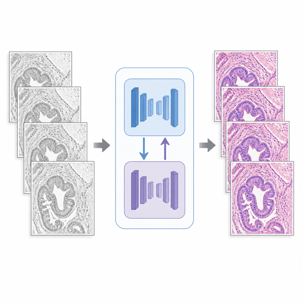
Can experts tell the difference
To probe whether these gains matter to human experts, the authors ran a blinded test with an experienced pathologist. The expert was shown mixed sets of real and virtual H&E and PAS-AB images, without knowing which was which, and asked to classify them. The pathologist’s accuracy hovered around chance level, just over 50 percent, with no statistically clear separation between real and virtual images. In follow-up experiments where virtual stains were fed into separate AI systems trained to classify tissue types and disease grades, models that received DGR-generated images performed as well as or better than those using only traditional stains, while several other virtual staining methods actually harmed performance.
What this means for future diagnoses
To a non-specialist, the main message is that careful AI design can turn virtual staining from a lab curiosity into a tool that works with messy, real-world data. By explicitly handling tissue misalignment instead of wishing it away, DGR creates digital stains that both look real to pathologists and preserve the subtle details needed for automated analysis. This could eventually allow hospitals to replace some chemical staining steps with fast, software-based ones, reducing costs, saving tissue, and shortening the time between biopsy and diagnosis while keeping diagnostic confidence intact.
Citation: Ma, J., Li, W., Li, J. et al. Generative AI for misalignment-resistant virtual staining to accelerate histopathology workflows. Nat Commun 17, 4494 (2026). https://doi.org/10.1038/s41467-026-71038-2
Keywords: virtual staining, histopathology, generative AI, digital pathology, tissue imaging