Clear Sky Science · en
Amplifying metabolic profiling of extracellular vesicle dynamics with ACTIVITY
Why tiny parcels in our lungs matter
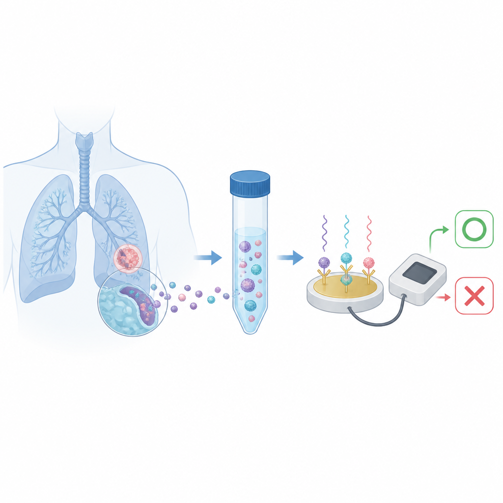
Doctors are eager to spot lung inflammation early, but current tools often rely on imaging that can be slow, costly, and expose patients to radiation. This study explores a very different idea: listening to the chemical activity of microscopic parcels released by immune cells in the lungs. By reading these signals from fluid collected during routine procedures, the researchers aim to turn these tiny parcels into a fast, sensitive indicator of pneumonia and its response to treatment.
Small messengers with hidden activity
Our cells constantly release nanosized bubbles called extracellular vesicles into body fluids such as blood and lung washings. These bubbles carry proteins and genetic material that mirror what is happening inside their parent cells, which has already made them attractive for blood tests known as liquid biopsies. The authors focus on vesicles shed by macrophages, immune cells that can adopt pro-inflammatory or anti-inflammatory roles. Instead of measuring how much of a protein is present inside the vesicles, they ask a more direct question: how active are the enzymes they carry, and can that activity reveal the state of inflammation inside the lung?
A chemical signature of angry immune cells
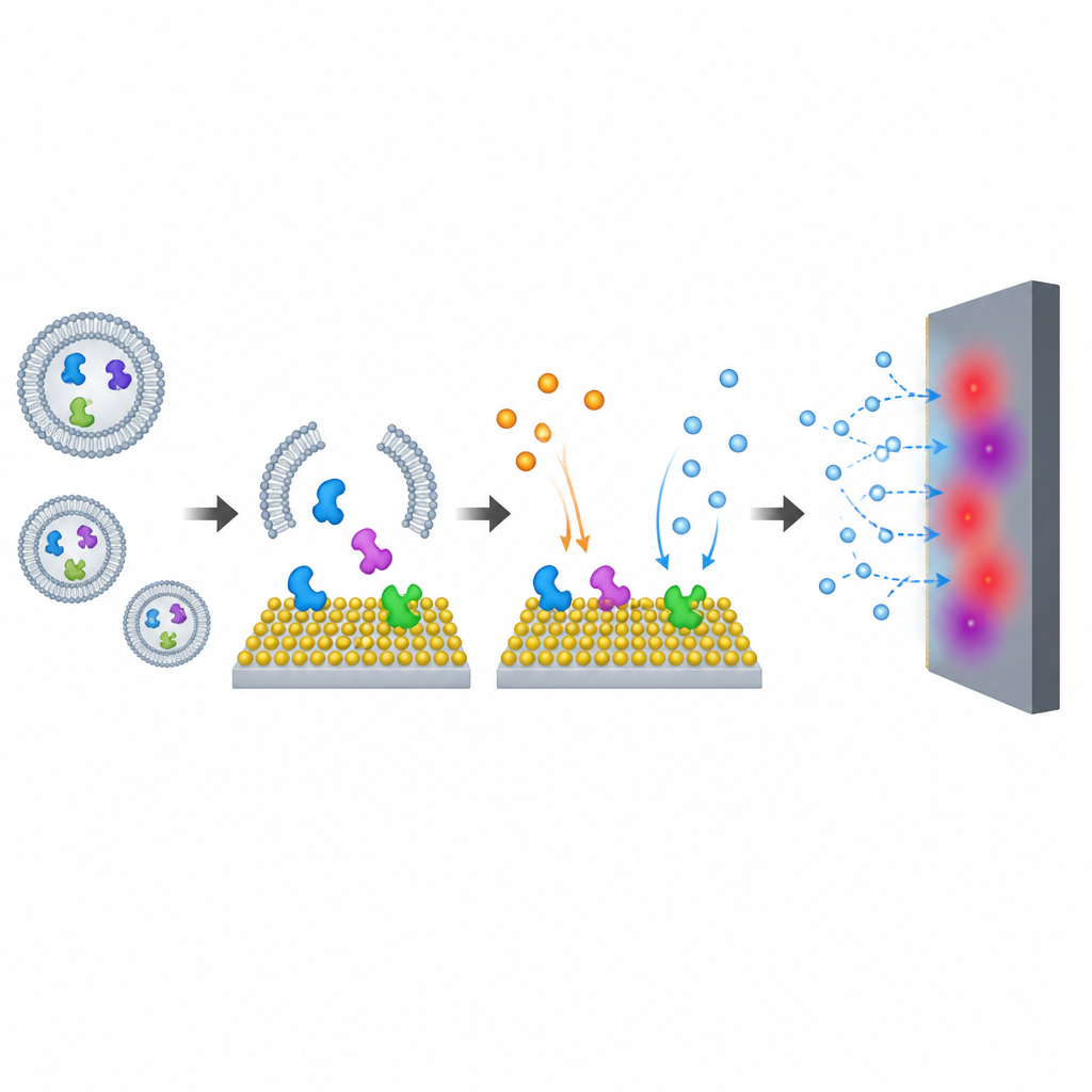
When macrophages shift into a strongly inflammatory mode, they ramp up an enzyme called inducible nitric oxide synthase, which converts a natural amino acid into nitric oxide, a reactive gas involved in immune defense. The team shows that this enzyme is packaged inside the vesicles released by such macrophages and remains stable for weeks when frozen. To detect its activity, they first break open the vesicles so the enzyme can work on its usual ingredients and generate nitric oxide. This gas then becomes the basis for an electrical signal that reflects how metabolically active the vesicles are and, by extension, how inflamed their parent cells are.
Boosting faint signals with smart materials
Because nitric oxide is produced in very small amounts, the researchers build a two-step amplification system they call ACTIVITY, short for Amplified Cascade-catalysis Testing for Vesicular Metabolic Activity. In the first step, the enzyme in the vesicles produces nitric oxide. In the second step, this gas is oxidized on an electrode coated with specially engineered quantum dots made from tungsten disulfide. By deliberately introducing defects into these nanomaterials, the team greatly improves their ability to convert nitric oxide into an electrical current. Careful testing shows that the most defective particles give the strongest and most reliable response, enabling detection of extremely low levels of active vesicles far beyond what standard antibody tests can achieve.
Reading lung inflammation from washed fluid
To see whether this approach has real-world value, the authors examined fluid collected from the lungs of pneumonia patients and healthy volunteers during bronchoalveolar lavage, a standard hospital procedure. They isolated vesicles from this fluid, normalized for vesicle quantity, and then measured their enzyme activity using the ACTIVITY system. Patients with pneumonia consistently showed higher vesicle activity than healthy controls. When the same samples were tested with a traditional protein-measuring kit, the separation between groups was much weaker. Statistical analysis revealed that the activity-based test outperformed both protein levels in vesicles and a common blood marker of inflammation in distinguishing sick from healthy individuals.
Watching treatment take effect in real time
The team also followed a small group of pneumonia patients over the course of therapy. In most cases, the metabolic activity of vesicles from their lung fluid dropped after treatment, in line with improvements seen on chest scans. This suggests that the method could help track how well a patient is responding without repeated radiation-based imaging. While the study notes that vesicles from several types of immune cells may contribute to the signal, macrophage-derived vesicles seem to dominate.
A new kind of liquid window into the lungs
Overall, the work suggests that it is not just what molecules are present inside extracellular vesicles that matters, but how chemically active they are. By turning that invisible enzyme work into a measurable electrical signal, the ACTIVITY method offers a sensitive, radiation-free way to gauge lung inflammation and monitor pneumonia treatment. With further refinement and portable devices, similar strategies could extend to other diseases where cell metabolism leaves a telltale trace in the tiny bubbles circulating through our body fluids.
Citation: Yu, RJ., Ma, WY., Xiao, HY. et al. Amplifying metabolic profiling of extracellular vesicle dynamics with ACTIVITY. Nat Commun 17, 4490 (2026). https://doi.org/10.1038/s41467-026-71030-w
Keywords: extracellular vesicles, pneumonia, lung inflammation, electrochemical sensor, nitric oxide