Clear Sky Science · en
An epithelial morphogenetic program for maximal urine concentration
How Kidneys Save Water
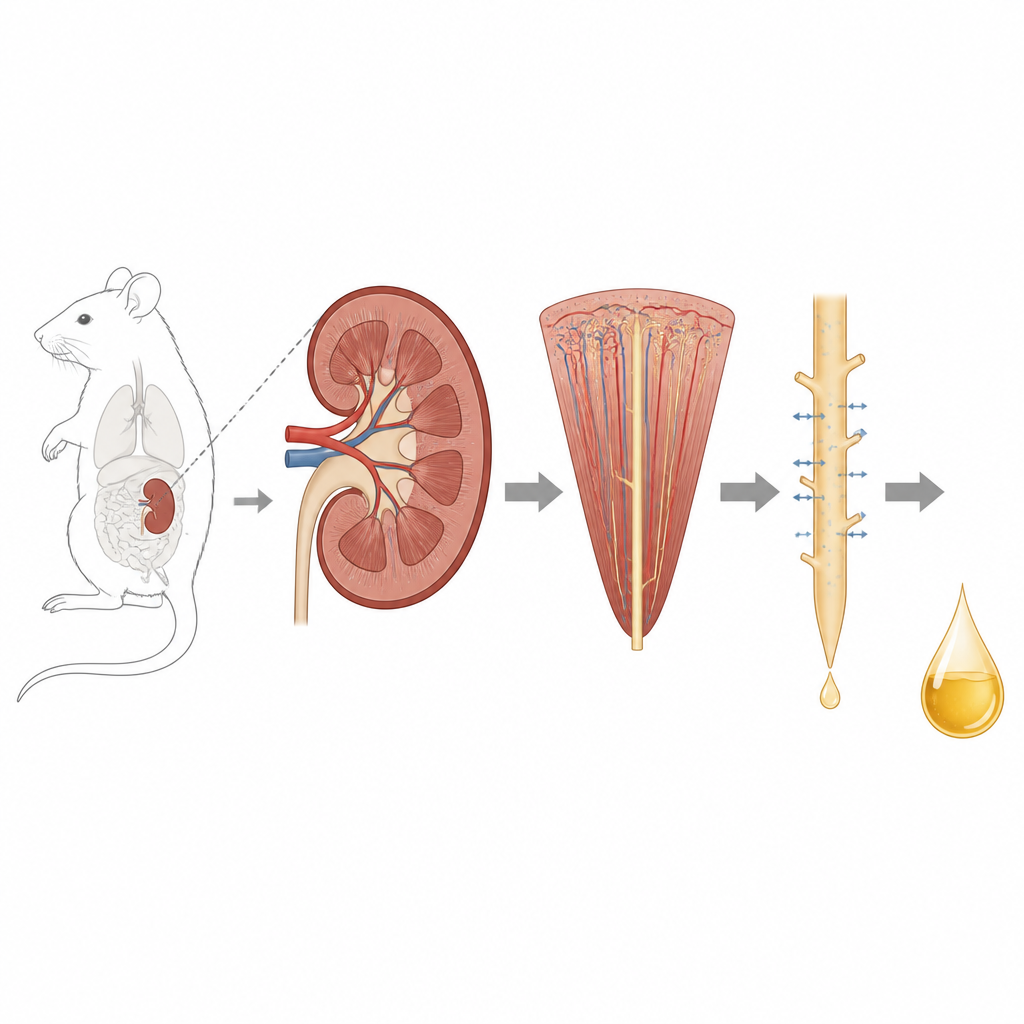
Mammals can survive in deserts and other dry places because their kidneys are experts at saving water while still removing waste. This study uncovers a hidden structural trick deep inside the kidney that helps create very salty conditions, allowing urine to become highly concentrated. By revealing how a specific group of kidney cells change their shape and how a single protein orchestrates this makeover, the work connects tiny changes at the cell level to a whole body ability that keeps animals alive when water is scarce.
A Hidden Zone Inside the Kidney
Each kidney is packed with tiny filtering units called nephrons, which twist and loop from the outer region into the inner zone. The deepest part, known as the inner medulla, is where urine reaches its highest concentration. In this region, a slim segment of the nephron called the ascending thin limb plays a unique role. Unlike other parts of the kidney that use energy-hungry pumps, this segment relies mostly on passive movement of dissolved salts. For decades, scientists suspected it was important for pulling salt out of the forming urine, but its exact structure and contribution to urine concentration remained mysterious, largely because it sits so deep in the organ and was hard to study with precise tools.
Finger-like Cell Projections Revealed
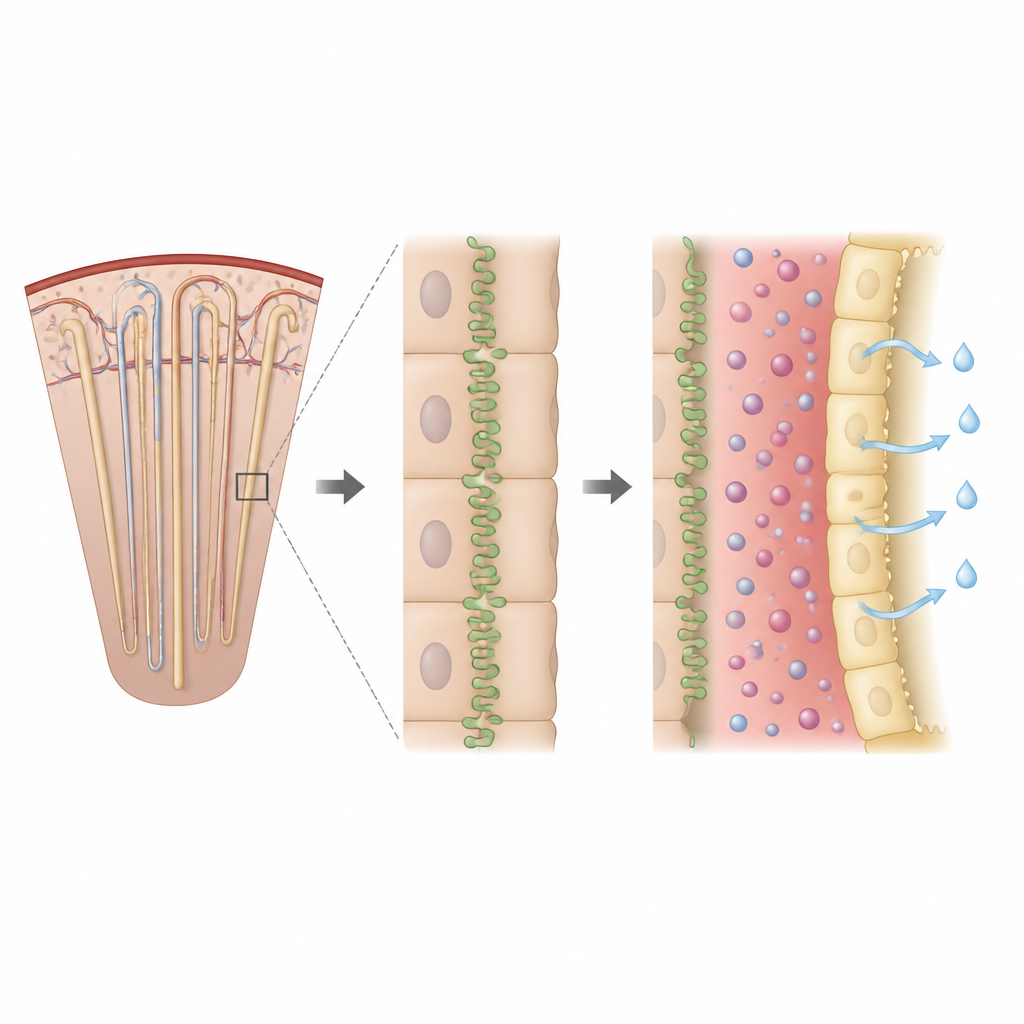
Using genetically tagged mice and advanced 3D microscopes, the researchers were able to label and visualize individual cells of this ascending thin limb in intact kidneys. Instead of forming smooth, flat borders, these cells displayed a striking sunburst-like shape. Their top edges extended numerous finger-like projections that wove between neighboring cells, greatly increasing the area where cells touch each other. These interlocking ridges developed after birth and continued to grow as the animals matured. Inside the projections, the team found key structural components such as actin, microtubules, and mitochondria, confirming that these were genuine extensions of the cell body rather than simple surface wrinkles.
The Role of a Single Junction Protein
To understand what builds this unusual architecture, the scientists turned to single-nucleus RNA sequencing, a technique that measures which genes are active in individual cells across the kidney. They identified a tight junction protein called claudin-10b as especially abundant in the ascending thin limb. This protein sits where neighboring cells meet and normally helps form a pathway for positively charged ions like sodium to slip between cells. In the inner medulla, it was concentrated precisely at the sites where the finger-like projections interlocked. When the team engineered kidney cell lines in a dish lacking several major tight junction proteins, adding claudin-10b alone was enough to restore a wavy, folded border between cells, suggesting that this protein actively sculpts the cell junctions.
From Cell Shape to Concentrated Urine
The researchers then removed the claudin-10b gene specifically from the ascending thin limb in mice, leaving the rest of the kidney intact. The affected cells lost their elaborate interlocking projections and showed much flatter surfaces, even though other junction proteins still reached the cell border. Functionally, these mice produced more dilute urine and larger urine volumes under normal conditions. When water was withheld for 24 hours, all mice concentrated their urine, but those lacking claudin-10b in this segment reached much lower maximum concentrations. Further experiments showed that claudin-10b must both stick to matching proteins on neighboring cells and bind to an internal scaffold protein called ZO1 to generate the projections. Mutations that disturbed adhesion or scaffold binding prevented projection formation, in both cultured cells and kidney tissue.
Why This Matters for Health and Evolution
These findings show that a single junction protein can serve two roles at once: shaping how kidney cells interlock and guiding how salt moves between them. Together, these actions help build a very salty environment in the inner medulla, which in turn allows water to be pulled back from the collecting ducts so urine becomes concentrated. By linking a specific cell shape program to whole-organ performance, the study explains how mammals achieve such high urine concentration and highlights a potential source of kidney problems when this system goes wrong. Rare human mutations in claudin-10 already cause salt imbalances and trouble concentrating urine, and this work suggests that damage to the ascending thin limb’s fine architecture may be an important part of that story.
Citation: Warshaw, J.N., Oh, S., Chaney, C.P. et al. An epithelial morphogenetic program for maximal urine concentration. Nat Commun 17, 4288 (2026). https://doi.org/10.1038/s41467-026-70938-7
Keywords: kidney, urine concentration, claudin-10b, renal tubule, osmotic gradient