Clear Sky Science · en
IgA autoantibodies promote inflammation, Th17 polarization and fibrotic responses in hidradenitis suppurativa
Painful skin disease under the microscope
Hidradenitis suppurativa is a chronic skin condition that causes painful lumps, abscesses, and scarring in areas such as the armpits and groin. Many people living with this disease are told that hair follicles get clogged and infected, but that explanation does not fully capture why the skin remains inflamed for years. This study looks inside diseased skin to track how a specific class of antibodies, called IgA, may help drive long lasting inflammation and scarring, offering a new way to think about this often misunderstood condition. 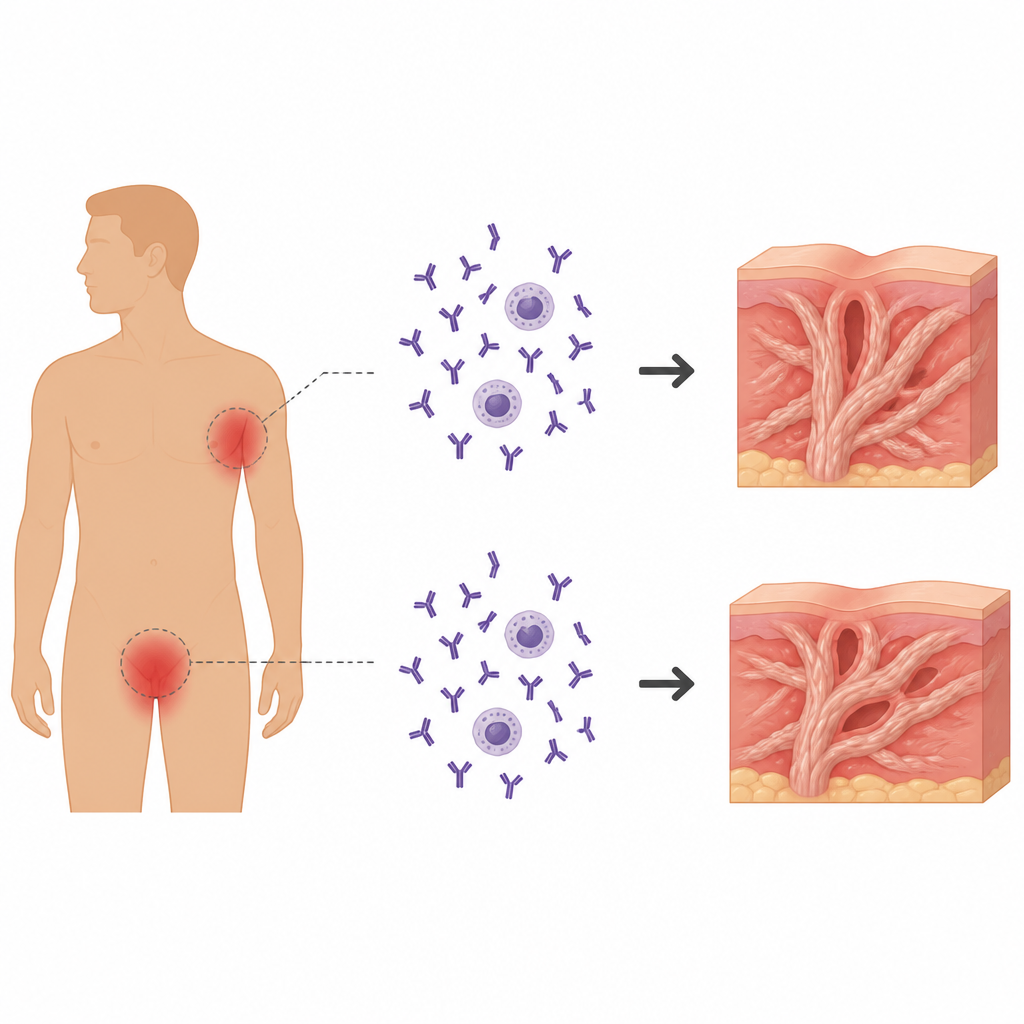
Antibodies show up in the wrong place
The researchers began by comparing skin samples from people with hidradenitis suppurativa to healthy skin. They found that IgA related genes and proteins were much higher in affected skin, but not in nearby normal looking skin or in the blood. Under the microscope, clusters of IgA producing cells sat next to groups of B cells in organized pockets that resembled miniature lymph nodes inside the skin. This pattern suggests that the diseased areas are not just passively inflamed but have become local factories that switch on B cells and churn out IgA antibodies right where the disease is most active.
Self targeting antibodies and links to symptoms
When the team analyzed what these IgA antibodies were sticking to, they discovered a broad mix of targets from the body itself, including parts of cell nuclei, the inside of cells, and structures outside cells such as collagen. Levels of many of these self directed IgA antibodies tracked with how severe a person’s disease was, how many tunnels and nodules they had, and with other health issues like diabetes or smoking. In contrast, IgA responses to common bacteria were not higher in patient skin, suggesting that these antibodies were not simply reacting to germs. Some IgA types were less common in patients taking certain anti inflammatory drugs, hinting that IgA patterns might one day help predict treatment responses.
How IgA stirs up immune cells and scar forming cells
To understand how IgA might worsen disease, the researchers recreated pieces of the skin environment in the lab. They showed that IgA from patient skin can coat proteins from skin cells and form immune clusters that are taken up by a type of sentry cell called a dendritic cell. These sentry cells then roused helper T cells to release strong inflammatory signals. Another set of experiments revealed that IgA attached to macrophages, a clean up cell type, pushing them to release powerful alarm molecules such as TNF, IL 6, and IL 1β. Fluids from these IgA activated macrophages were able to steer fresh T cells toward a Th17 like state, a flavor of immune response already suspected to be important in this disease. 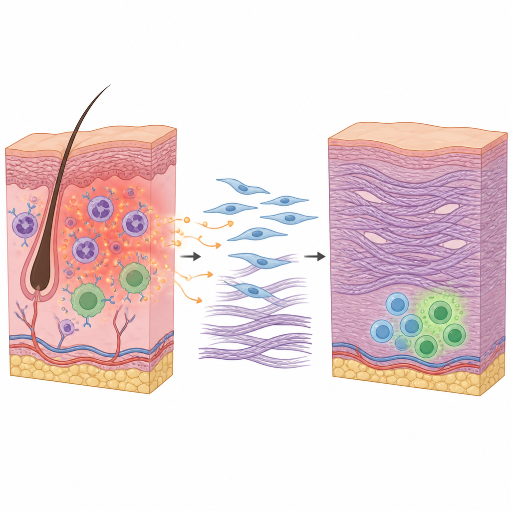
A vicious loop of traps, antibodies, and scarring
The study also uncovered a feedback loop involving structures called neutrophil extracellular traps, sticky webs released by white blood cells. Patient IgA recognized these traps and even encouraged more of them to form. When traps bound to IgA were presented to macrophages, the cells secreted CCL18, a signal known to push skin fibroblasts to make excess collagen. Fibroblasts exposed to cues from these immune clusters switched on genes linked to type I interferon responses, inflammatory messengers, and tissue stiffening. When fibroblasts were directly exposed to trap IgA clusters, they began to express molecules that attract and hold immune cells, and factors that support B cells, suggesting that scar forming cells themselves help maintain the chronic immune hub inside the skin.
What this means for people with this disease
Taken together, the work paints hidradenitis suppurativa as more than a problem of blocked follicles or surface infection. Instead, the diseased skin behaves like an active immune organ where IgA producing B cells, antibodies, white blood cells, and fibroblasts talk to one another in a self reinforcing cycle. IgA autoantibodies sit at the center of this loop, helping to sustain inflammation, recruit aggressive T cells, and drive scarring. Understanding this network may open doors to new treatments that target IgA, its receptors, or the skin based immune hubs that support it, with the long term goal of easing pain, preventing new lesions, and limiting permanent tissue damage.
Citation: Carmona-Rivera, C., O’Neil, L.J., Patino-Martinez, E. et al. IgA autoantibodies promote inflammation, Th17 polarization and fibrotic responses in hidradenitis suppurativa. Nat Commun 17, 4469 (2026). https://doi.org/10.1038/s41467-026-70883-5
Keywords: hidradenitis suppurativa, IgA autoantibodies, chronic skin inflammation, fibrosis, Th17 immune response