Clear Sky Science · en
A prospective multicenter trial of deep learning auto-segmentation for organs at risk in thoracic radiotherapy
Why this matters for people with chest cancers
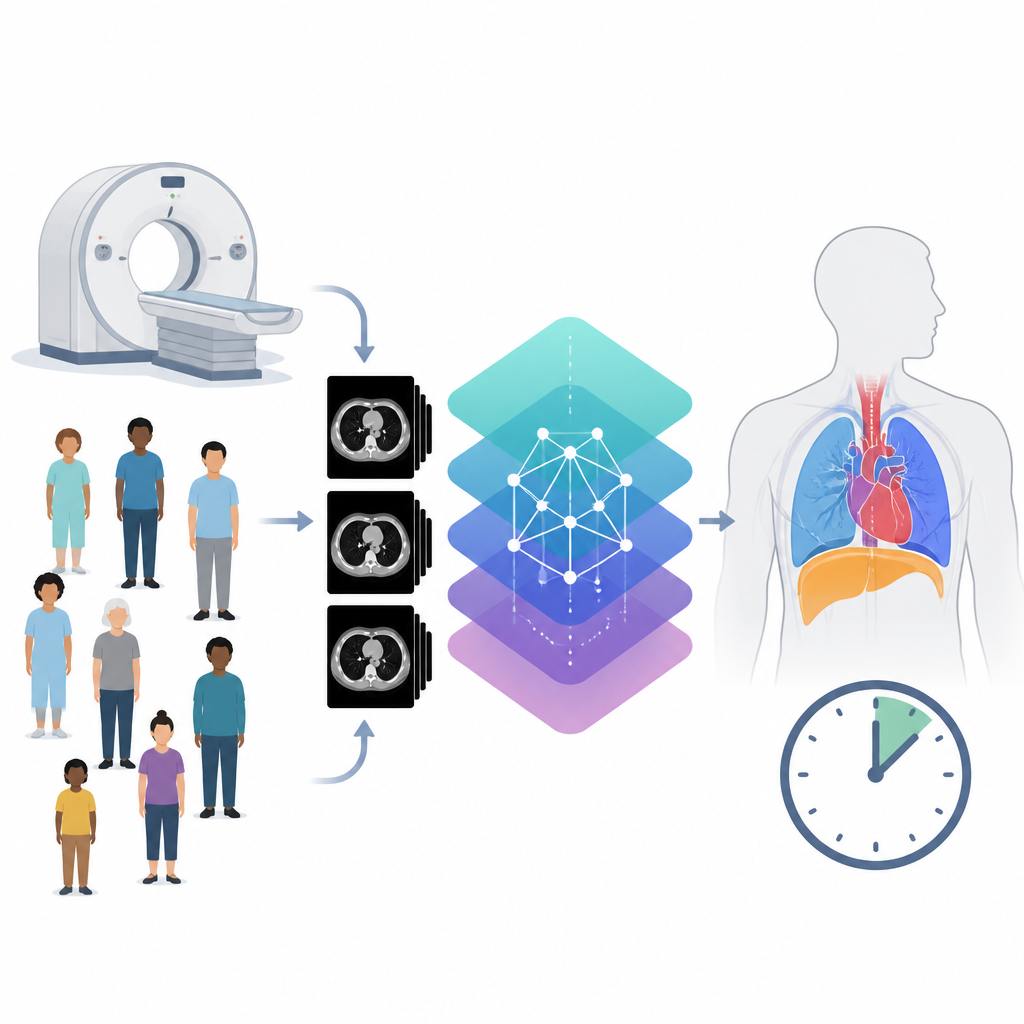
For people facing lung, breast, or esophageal cancer, radiation therapy is often a key part of treatment. But before any beam is turned on, doctors must carefully outline vulnerable organs in the chest so radiation can be aimed at tumors while sparing healthy tissue. This study tested whether a form of artificial intelligence, used together with physicians, can make that outlining faster, more accurate, and more consistent across hospitals.
How chest scans guide radiation treatment
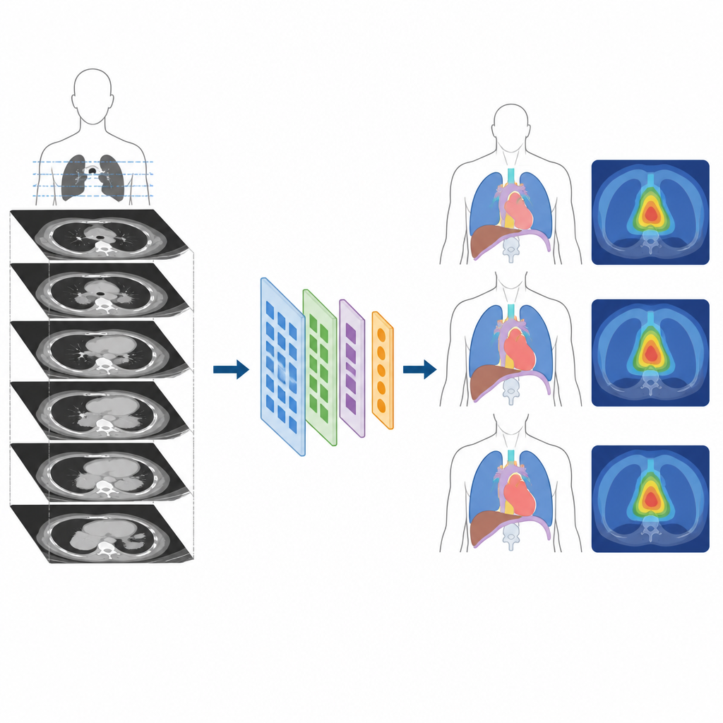
Radiation plans are built on detailed CT scans of the chest. On these scans, doctors draw the borders of organs that could be harmed by radiation, such as the lungs, heart, spinal cord, and esophagus. These are called organs at risk. Doing this slice by slice is slow and depends heavily on each doctor’s experience and local training, which can lead to differences in how organs are drawn and in how much radiation they appear to receive.
What the researchers set out to test
The team evaluated a deep learning system named iCurveE that automatically outlines eleven key organs in the chest. Instead of only checking how the computer performed on old data, they organized a prospective trial across five hospitals in China. Five hundred patients with lung, breast, or esophageal cancer had their planning CT scans outlined in three ways: fully by hand, by the AI alone, and by physicians who started from the AI outlines and then edited them. In total, 37 radiation doctors of varying experience levels contributed almost 2,500 full organ sets.
How well human plus AI worked together
To compare methods, the researchers used measures of how closely each set of organ borders matched a carefully agreed-upon expert reference. They also recorded how long each approach took. On average, physician-edited AI outlines matched the expert reference better than outlines drawn fully by hand and slightly better than the AI alone. A key distance measure of border mismatch was lower with AI assistance than with manual drawing, and a standard overlap score was higher. At the same time, using AI as a starting point cut the median outlining time from 55 minutes to 10 minutes, an improvement of more than 80 percent, and this time saving held up across all hospitals and cancer types.
Consistency across hospitals and experience levels
One concern in cancer care is that patients at smaller or less well-resourced hospitals may not receive the same quality of planning as those at major centers. In this study, manual outlining varied significantly between hospitals and between less-experienced and more-experienced doctors. When physicians worked from AI-generated outlines, those differences shrank. The gap in accuracy and time between centers narrowed, and less-experienced doctors became more similar to their senior colleagues. The combination of AI and human review also led to smaller differences in the calculated radiation doses to organs, suggesting that better outlining can translate into more reliable estimates of treatment risk.
What this means for future cancer care
This trial shows that when doctors carefully review and adjust AI-generated contours, they can draw chest organs more quickly and with greater accuracy than by hand alone, while reducing differences between hospitals and between physicians. For patients, this could mean safer and more consistent radiation plans, regardless of where they are treated. The study also highlights that AI is a tool rather than a replacement for clinicians: human oversight remains essential to catch unusual anatomy, imaging problems, and model errors. As similar systems are tested and refined in other body regions and in outcome-focused trials, human–AI teamwork may become a routine part of planning radiation therapy.
Citation: Niu, G., Guan, Y., Zhang, Y. et al. A prospective multicenter trial of deep learning auto-segmentation for organs at risk in thoracic radiotherapy. Nat Commun 17, 4633 (2026). https://doi.org/10.1038/s41467-026-70863-9
Keywords: thoracic radiotherapy, auto segmentation, deep learning, organs at risk, medical imaging AI