Clear Sky Science · en
Major human schistosome species express different glycans with immunological and diagnostic implications
Why sugar coatings on parasites matter
Many disease-causing microbes hide behind sugar coats on their surfaces. These sugar patterns help them slip past our defenses, but they can also act as flags that our immune system learns to recognize. This study looks at the sugar coats of two major worm species that cause schistosomiasis in humans and shows that, although closely related, they decorate themselves with surprisingly different sugars. Those differences help explain how the body responds to each parasite and point to new ways to detect infections more accurately.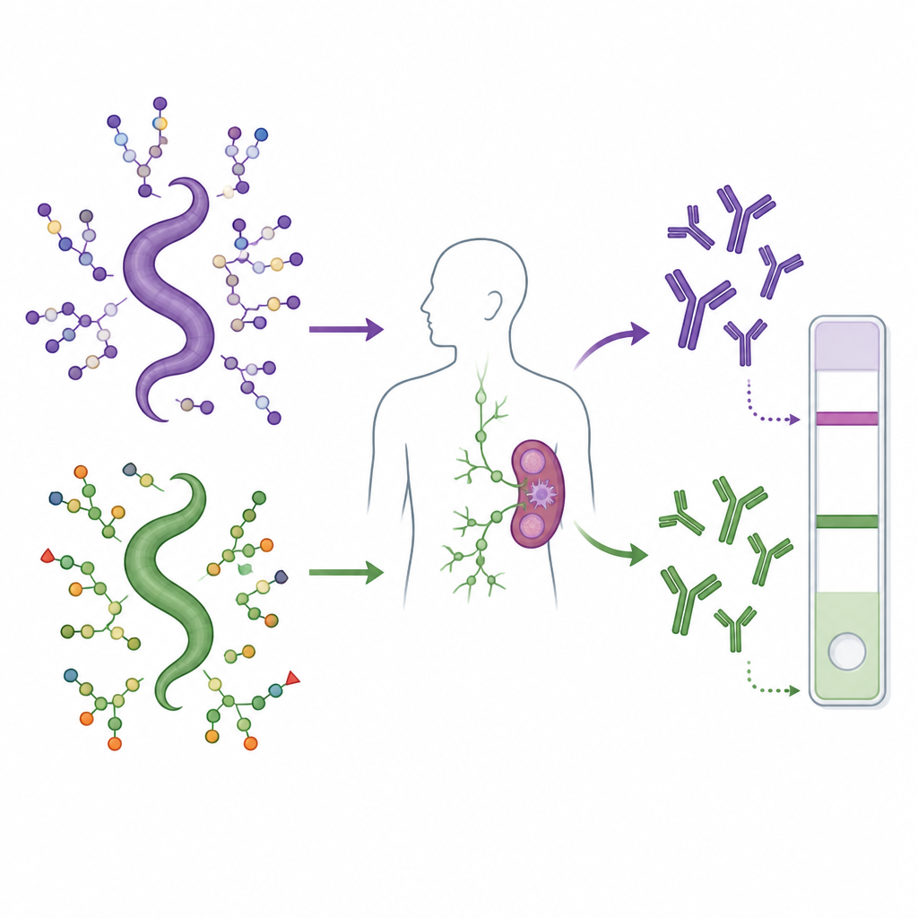
Two similar worms, two different diseases
Schistosomiasis is a chronic parasitic disease that affects hundreds of millions of people, mostly in tropical regions with poor access to clean water. One worm species, Schistosoma mansoni, mainly damages the intestines and liver. Another, Schistosoma haematobium, targets the urinary and genital organs and is linked to bladder cancer. Current control relies on a single drug and basic sanitation, and there is no vaccine. To better fight these infections, scientists need to understand the basic biology of the worms, including the sugar structures they present to the human immune system.
Mapping the sugar coats across worm life stages
The researchers compared the sugars attached to fats and proteins in different life stages of S. haematobium and contrasted them with what is known for S. mansoni. They focused on three key stages: free-swimming larvae that infect humans, adult worms living in blood vessels, and eggs that lodge in tissues and trigger disease. Using advanced mass spectrometry and specialized separation techniques, they pieced together which building blocks make up these sugars and how they are linked. This revealed complex, often branched chains with many variants, rather like decoding a set of barcodes that shift as the parasite develops.
Unique sugar signatures in the bladder parasite
One of the striking findings is that S. haematobium builds many of its fat-linked sugars on a three-sugar core that differs from the two-sugar core used by S. mansoni. In addition, S. haematobium uses fewer “fucose” decorations, a type of sugar that is heavily used by S. mansoni and is known to strongly stimulate the immune system. Instead, S. haematobium eggs carry many sugars that include glucuronic acid, an acidic sugar that gives the chains a negative charge. These acidic units can sit at or just beneath the tips of the chains, in positions where immune molecules are likely to notice them. Such structures are rare in human sugars but common in several parasites, making them good candidates for species-specific markers.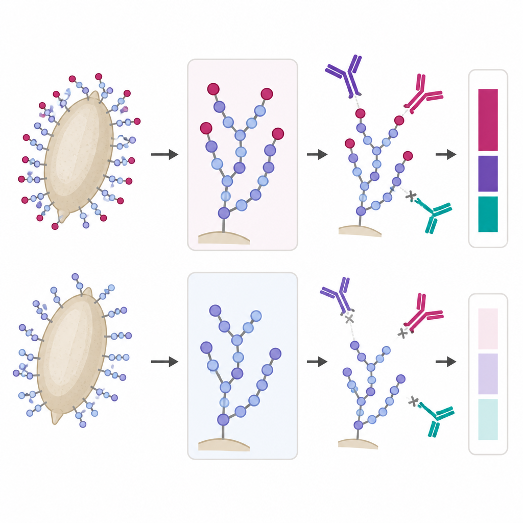
How children’s antibodies read these sugar codes
To see how the human immune system responds to these sugar designs, the team printed purified parasite sugars onto glass slides, creating a kind of sugar microchip. They then exposed this array to blood samples from children infected with either S. haematobium or S. mansoni, as well as from uninfected people. Both groups of infected children produced antibodies that strongly recognized many worm sugars, especially those with several fucose units. But there was a clear difference for the acidic sugars: children infected with S. haematobium had much higher levels of IgG antibodies that bound sugars containing glucuronic acid from S. haematobium eggs, while this response was weak in children infected with S. mansoni. When the scientists chemically removed fucose from the sugars, binding by these antibodies hardly changed, showing that it was the acidic part that really mattered here.
From basic sugar maps to better tests
This work shows that the two main human schistosome species dress themselves in notably different sugar patterns. The bladder parasite, S. haematobium, relies less on dense fucose decoration and more on acidic sugars that stand out to the immune system. These distinctive sugar motifs act as clear signals that the body responds to with specific antibodies, especially in the case of S. haematobium infection. In practical terms, these findings mean that carefully chosen sugars from S. haematobium eggs could be turned into blood tests that distinguish between worm species and help track this neglected disease more precisely.
Citation: Petralia, L.M.C., van Diepen, A., Zhang, T. et al. Major human schistosome species express different glycans with immunological and diagnostic implications. Nat Commun 17, 4312 (2026). https://doi.org/10.1038/s41467-026-70850-0
Keywords: schistosomiasis, parasite glycans, Schistosoma haematobium, immune response, diagnostics