Clear Sky Science · en
Optically driven control of mechanochemistry and fusion dynamics of biomolecular condensates via thymine dimerization
Light that stiffens cellular droplets
Inside living cells, many molecules gather into tiny liquid droplets that help organize life’s chemistry. This study shows that ultraviolet (UV) light can act as an invisible dial to tune how these droplets flow, fuse, and hold their contents, all without adding new ingredients. The work links everyday sunlight-driven damage to DNA with the physics of soft materials, and even hints at how early “protocells” on ancient Earth might have coped with harsh radiation.
Tiny droplets that act like rooms
Cells contain biomolecular condensates, droplet-like compartments formed when certain proteins and nucleic acids separate from the surrounding fluid, much like oil in water. These droplets are usually soft and liquid-like, letting molecules move in and out and react efficiently. Their internal consistency, however, can vary from runny to gel-like or even solid. That consistency strongly affects how molecules travel and react inside them, which in turn shapes cell behavior and may influence diseases linked to protein aggregation. Until now, most ways of changing droplet mechanics involved adding extra chemicals or changing salt levels, which also alters the makeup of the droplets themselves.
Using UV light to reshape DNA-based droplets
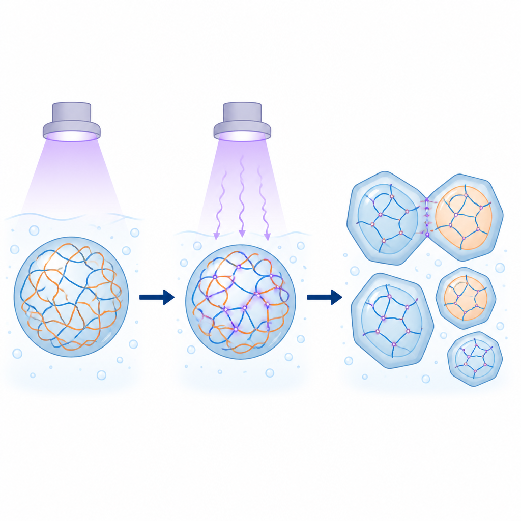
The researchers created a simple model of these condensates using short strands of DNA made of thymine bases and a positively charged peptide, poly-L-lysine. These ingredients spontaneously formed microscopic droplets under suitable salt conditions. The team then exposed the samples to UVC light, a strong form of UV that makes nearby thymine bases in DNA bond together, forming tiny “dimers.” Using optical microscopy, spectroscopy, and antibody staining, they confirmed that these dimers formed inside the droplets. Crucially, UV changed the size and shape of the droplets: longer exposure led to more elongated shapes, a higher chance of droplets sticking as pairs or clusters, and a shift in the statistics of droplet sizes, all signs that the material was becoming more rigid and less freely flowing.
Measuring how droplets harden and how they fuse
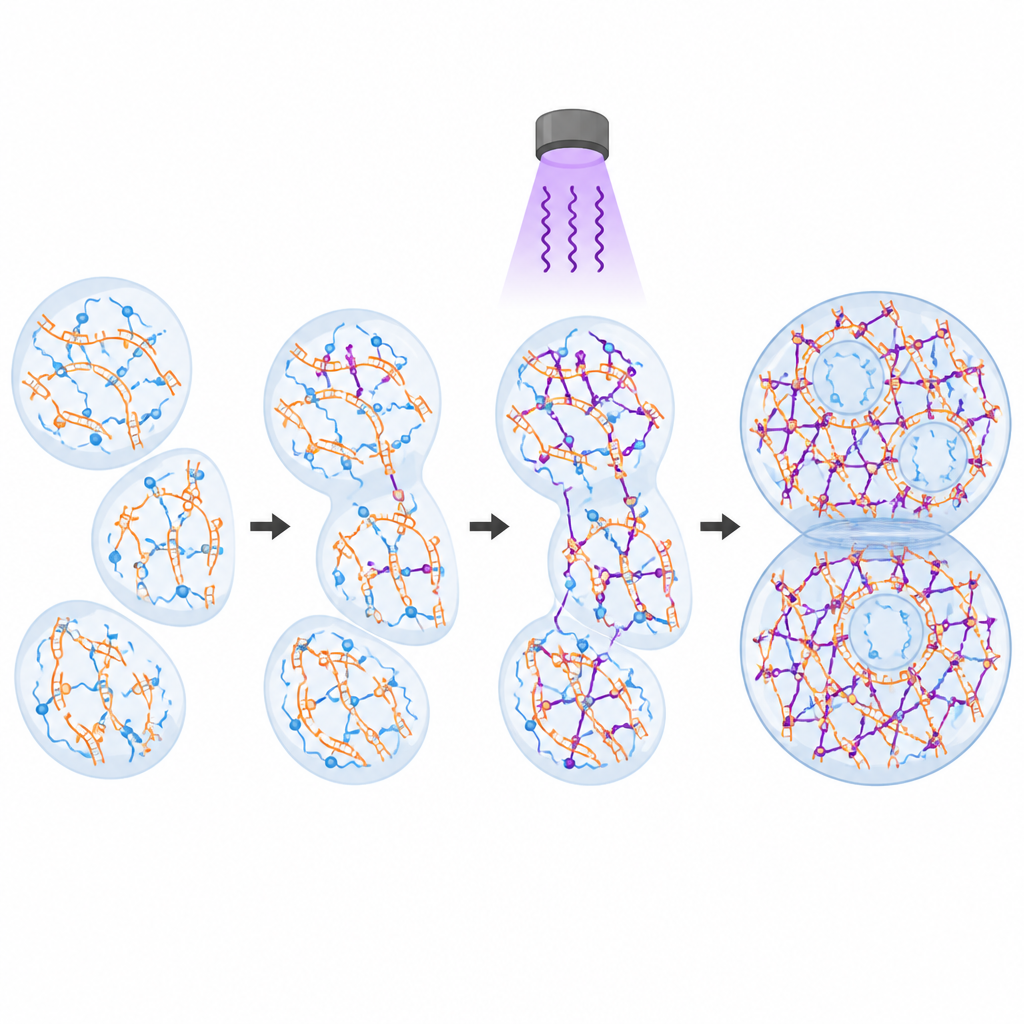
To probe how UV affected droplet mechanics, the team used scanning probe microscopy, a technique in which a tiny cantilever gently presses and oscillates against a single droplet to measure how it resists deformation. Before irradiation, droplets behaved like simple liquids, with energy mostly lost as viscous flow. After moderate UV exposure, the droplets showed a clear transition toward solid-like behavior: both elastic and viscous responses increased sharply, and at higher frequencies the droplets behaved more like soft gels than runny fluids. Stronger UV treatment created an even stiffer and more heterogeneous material. By adapting the same tool, the researchers developed an assay to bring two droplets into contact and record the forces during fusion. Untreated droplets coalesced quickly, dominated by surface tension, whereas UV-treated droplets fused slowly, with weaker adhesion and force traces that revealed a strong role for internal viscoelastic resistance.
When and where light strikes makes a difference
The timing of UV exposure turned out to be crucial. If DNA was irradiated before mixing with the peptide, droplets still formed but were smaller and remained largely liquid-like, consistent with bonds forming mainly within single DNA strands. When the mixture was irradiated immediately after mixing, instead of droplets the system produced an extended network of aggregates, suggesting abundant bonds between different chains. When UV was applied after droplets had already formed, it selectively strengthened the existing droplets, increasing crosslinks between chains within the dense interior. These crosslinks slowed molecular exchange, as shown by reduced uptake of fluorescent DNA and almost no recovery in fluorescence after photobleaching. A simple model captured how the balance between bonds within and between chains shapes both stiffness and fusion forces.
Stable droplets and clues to early life
UV-treated droplets proved remarkably robust to extreme environmental changes. When the surrounding liquid was suddenly replaced with pure water or very salty solution, conditions that normally dissolve such condensates, the droplets persisted. In low salt, they even developed internal dilute pockets, revealing a form of stable compartmentalization within a single droplet. This suggests that UV-driven crosslinking can lock in droplet structure and create internal “rooms” that respond slowly to outside shifts. The authors propose that on early Earth, such light-hardened condensates might have helped protect primitive genetic material while still allowing useful chemistry, and that similar principles could be harnessed today to build light-programmable soft materials and synthetic organelles.
Citation: Sheikhhassani, V., Wong, F.H.K., Bonn, D. et al. Optically driven control of mechanochemistry and fusion dynamics of biomolecular condensates via thymine dimerization. Nat Commun 17, 4436 (2026). https://doi.org/10.1038/s41467-026-70757-w
Keywords: biomolecular condensates, UV light, thymine dimers, phase separation, protocells