Clear Sky Science · en
RF-SIRF reveals a replication stress-specific epigenetic code by spatio-temporal mapping of reversed forks
Why tiny DNA traffic jams matter
Every time a cell copies its DNA, it must faithfully duplicate billions of letters without tearing or stalling. When this copying process runs into trouble, it creates molecular traffic jams that can lead to aging, cancer, and failed responses to cancer therapy. This study introduces a new way to watch those traffic jams form and resolve inside individual cells, revealing that they carry a distinctive chemical signature that helps cells decide how to respond to stress.
New way to see DNA stalls inside cells
When DNA copying slows or stops, the fork-like structures that separate the two strands can bend backward into a four-armed shape called a reversed fork. Until now, scientists could reliably see these shapes only by using high-powered electron microscopes on purified DNA, a demanding method that loses the context of the living cell. The authors built on a microscopy technique called SIRF to create RF-SIRF, which uses brief labeling of freshly made DNA and a proximity-based fluorescent reaction to light up places where the new DNA strands are pressed closely together, as happens at reversed forks. By comparing this signal to a separate measure of how much new DNA was made, they can quantitatively detect reversed forks in single cells.
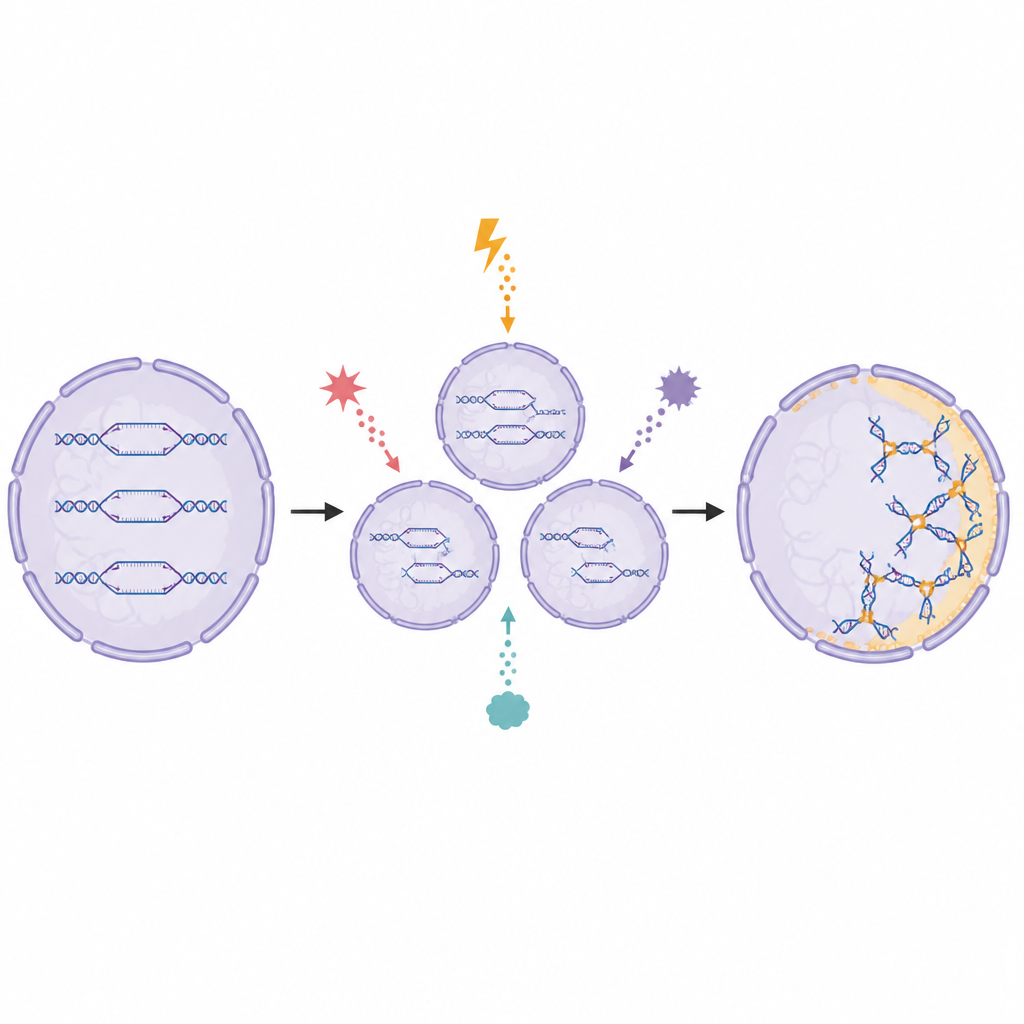
How cells respond to different kinds of stress
The team tested RF-SIRF under a variety of gentle DNA stresses that are known to trigger fork reversal, including several drugs and oxidizing molecules. Even after only 15 minutes of treatment, RF-SIRF showed a clear rise in reversed fork signal for all these agents, while the amount of newly made DNA changed far less. This pattern indicates that the assay responds mainly to changes in fork shape rather than simple slowing of DNA copying. When the researchers blocked key enzymes that are required to bend forks backward, the RF-SIRF signal dropped markedly, confirming that the bright spots indeed mark reversed forks, not other unusual DNA structures.
Where and when the forks stall
Because RF-SIRF works in intact cells, it can reveal when during the cell cycle and where inside the nucleus these structures appear. By combining the new signal with markers that show early, middle, or late stages of DNA copying, the authors found that reversed forks are most abundant in early and mid S-phase, when cells first embark on duplication of their genomes. Surprisingly, many of these signals form ring-like patterns at the outer edge of the nucleus, near the nuclear shell, while ordinary replication continues throughout the interior. In late S-phase, reversed forks still appear but are more evenly scattered, suggesting that different regions of the genome may experience and manage stress in distinct nuclear zones.
A special chemical code on stressed DNA
DNA in cells is wrapped around proteins and decorated with small chemical tags, creating chromatin. These tags tell the cell which regions are active, silent, or under repair. Using RF-SIRF combined with antibodies that recognize specific tags, the researchers found that reversed forks are coated with a unique blend of marks that differs from those seen on genes being switched on or off. A classic “silent” mark (H3K9me3) and an “active” mark (H3K4me3) both accumulate at reversed forks, while another active mark (H4K16ac) is depleted. These combinations depend on fork reversal enzymes and on a tagging factor called PTIP, implying that cells deliberately write a mixed signal on stressed forks. This mixed pattern helps explain why certain repair proteins are drawn to these sites while others are kept away.
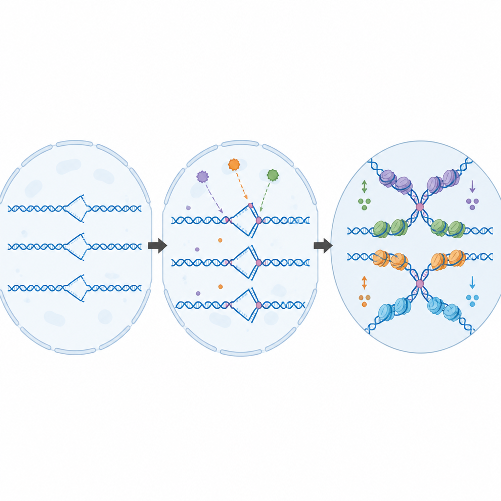
What this means for health and disease
Together, the findings show that reversed forks are not just passive byproducts of stalled DNA copying, but carefully managed structures that carry their own epigenetic code, especially during the earliest stages of genome duplication at the nuclear edge. RF-SIRF makes it possible to map these stressed sites and their protein partners in single cells with standard microscopes, opening the door to studies of how replication stress shapes development, aging, blood formation, and the response of tumors to chemotherapy. For a lay reader, the key message is that cells mark dangerous DNA traffic jams with a distinct chemical language, and this new method finally lets scientists read that language in place.
Citation: Roy, S., Fimreite, M.M., Chen, Y. et al. RF-SIRF reveals a replication stress-specific epigenetic code by spatio-temporal mapping of reversed forks. Nat Commun 17, 4302 (2026). https://doi.org/10.1038/s41467-026-70716-5
Keywords: DNA replication stress, reversed replication forks, chromatin marks, epigenetic code, cancer therapy response