Clear Sky Science · en
DEUP1 functions as a scaffold for basal foot integrity and planar polarity in multiciliated cells
How Tiny Hairs Help the Brain’s Fluid Flow
Inside the brain’s ventricles, a clear liquid called cerebrospinal fluid continuously washes over nerve tissue. This flow is powered by countless microscopic “hairs,” or cilia, that beat in unison on specialized cells. This article explores how a little‑known protein, DEUP1, quietly keeps these hairlike structures aligned and working together over a lifetime—and what happens when that support fails.
Microscopic Brushes that Keep Fluid Moving
Many surfaces in our bodies are lined with multiciliated cells—cells bearing dozens to hundreds of cilia that beat rhythmically to move fluid. In the brain, multiciliated ependymal cells line the ventricles and help propel cerebrospinal fluid, which carries nutrients, clears waste, and shapes how new neurons migrate. Each cilium is rooted in the cell by a basal body, much like a flagpole in the ground. A small, angled projection called the basal foot sticks out from each basal body and points in the direction of the ciliary beat. When thousands of basal feet share the same orientation, cilia beat in a coordinated wave and fluid flows in a single, efficient direction. 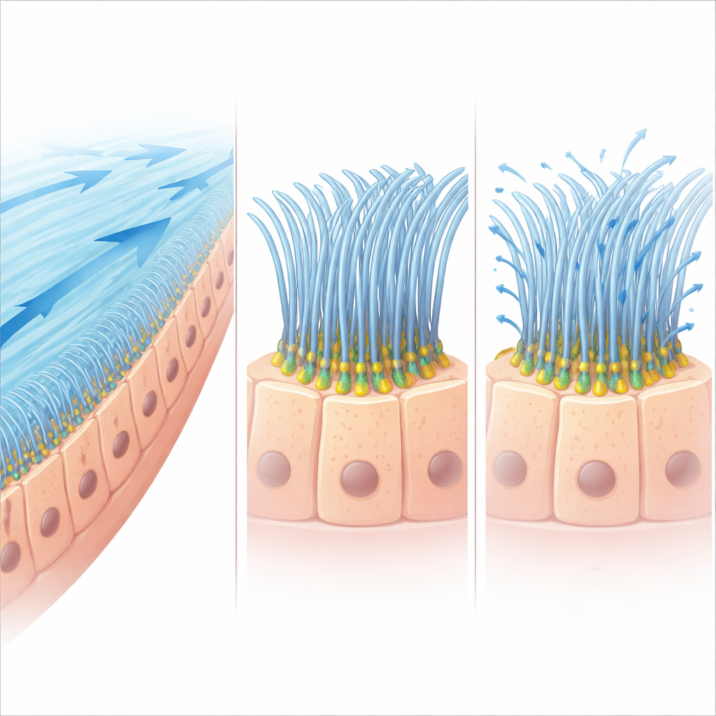
A Surprising New Job for a Known Protein
DEUP1 was originally famous for something else: helping cells rapidly manufacture the many basal bodies needed to build motile cilia. Because of this, scientists assumed that losing DEUP1 would prevent multiciliated cells from forming enough cilia in the first place. But earlier work in mice showed a surprise—animals completely lacking DEUP1 still made normal numbers of basal bodies and cilia and seemed healthy. This raised a puzzle: if DEUP1 is not essential for building cilia, what is it doing in these cells?
DEUP1 as a Structural Brace in the Basal Foot
Using high‑resolution light and electron microscopy, the authors mapped where DEUP1 sits in fully formed ependymal cells. Rather than clustering in factories for new basal bodies, DEUP1 was found embedded in the basal foot itself, next to another structural protein called CNTRL. Together, these proteins occupy a middle “tier” of the basal foot between regions that anchor it to the basal body and regions that grab onto the cell’s internal scaffold of microtubules. When the team knocked out DEUP1 in mice, this basal foot structure shrank: key components moved closer to the basal body, and the overall volume of the basal foot cone decreased. Over time, the upper portion of the basal foot, where microtubules attach, appeared to collapse toward the cell surface. 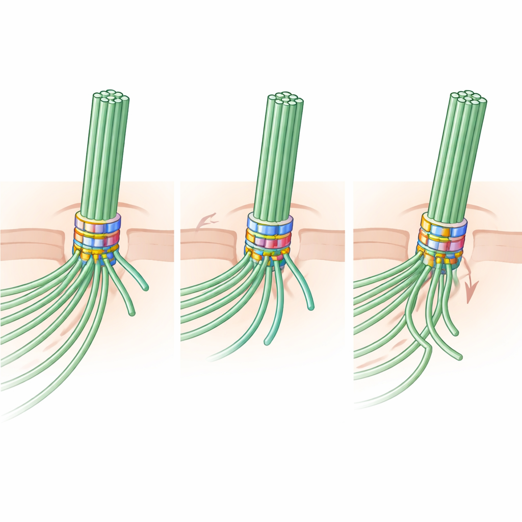
From Microscopic Misalignment to Sluggish Fluid Flow
The shape of the basal foot matters because it encodes the direction in which each cilium will beat—a property known as rotational planar polarity. In healthy young mice, the basal feet of neighboring basal bodies all point in nearly the same direction, and fluid flows smoothly along the ventricular wall. In mice lacking DEUP1, basal feet become misaligned, and their angles are much more scattered. Tiny tracer beads placed on the ependymal surface revealed that cerebrospinal fluid still flowed, but more slowly and less straight than in normal animals. Early on, the number of basal bodies and cilia per cell was largely unchanged; the main problem was loss of coordinated orientation rather than outright failure to make cilia.
Long-Term Wear, Tear, and Evolutionary Conservation
As DEUP1‑deficient mice aged, the consequences became more severe. The mechanically weakened basal feet appeared to offer poor anchoring as cilia beat against the constant drag of fluid. In old animals, both basal bodies and cilia were progressively lost, especially in regions exposed to strong fluid shear near the brain’s fluid outflow points. Some aged knockout mice developed enlarged brain ventricles, consistent with less efficient fluid clearance. To test whether this role for DEUP1 is unique to mammals, the authors turned to the skin of frog embryos, another classic multiciliated system. There, DEUP1 again localized to the basal foot, and blocking its production disrupted basal foot orientation and, later, the stability of basal bodies—showing that this scaffolding function is conserved across vertebrates.
Why This Matters for Brain Health
Overall, the study recasts DEUP1 not as a simple builder of new cilia, but as a long‑term structural brace for the basal foot. By helping to maintain the size and shape of this tiny projection, DEUP1 keeps cilia pointing in the same direction and beating in coordinated waves, which preserves robust cerebrospinal fluid flow and protects the underlying cell skeleton from chronic mechanical stress. Over decades of life, this microscopic alignment may be one of the quiet safeguards that prevents subtle fluid‑flow problems from contributing to brain aging and disease.
Citation: Lee, H., Lee, J., Shin, M. et al. DEUP1 functions as a scaffold for basal foot integrity and planar polarity in multiciliated cells. Nat Commun 17, 3875 (2026). https://doi.org/10.1038/s41467-026-70661-3
Keywords: multiciliated cells, cerebrospinal fluid flow, basal foot, ciliary polarity, DEUP1 protein