Clear Sky Science · en
Structural basis and regulation of GSDME pore formation
How Cells Turn a Quiet Signal into a Fiery Death
Our immune system sometimes defends us by allowing damaged or infected cells to self-destruct in a dramatic, explosive way that alarms nearby defenses. This process, called pyroptosis, punches holes in cell membranes so that danger signals can spill out and rally the immune system against infections or tumors. The protein GSDME has emerged as a key player in this fiery form of cell death, especially in cancer cells treated with chemotherapy, but until now scientists did not fully understand how it assembles those membrane holes or how the cell controls its power.
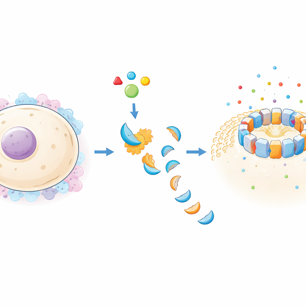
A Protein That Builds Deadly Doorways
GSDME belongs to a family of proteins that, once activated, form large rings in cell membranes. These rings act like wide-open doorways that let water and molecules rush in and out, rupturing the cell. Using cryo-electron microscopy, a technique that images frozen molecules at near-atomic resolution, the researchers solved detailed 3D structures of human GSDME pores. They found that each pore is built from 27 or 28 copies of the GSDME active fragment, arranged in a near-perfect circle that spans the cell membrane. Each copy looks like a tiny hand: a compact “palm” sits inside the cell, while long “fingers” reach through the membrane to form a continuous tunnel.
What Makes GSDME’s Holes Special
Although GSDME resembles its gasdermin relatives, its pores have distinctive features that may shape how and where it acts in the body. Its membrane-spanning barrel is longer than those of other family members, allowing it to cross a slightly thicker layer of fat molecules. The researchers also discovered that the inside of the GSDME pore carries a negative charge, which would help positively charged immune molecules—such as certain cytokines—escape the cell more easily. Compared with related proteins, GSDME’s “hand” sits closer to the membrane surface, creating a more compact footprint that may influence which cellular membranes it prefers, such as those enriched in specific lipids found in mitochondria or tumor cells.
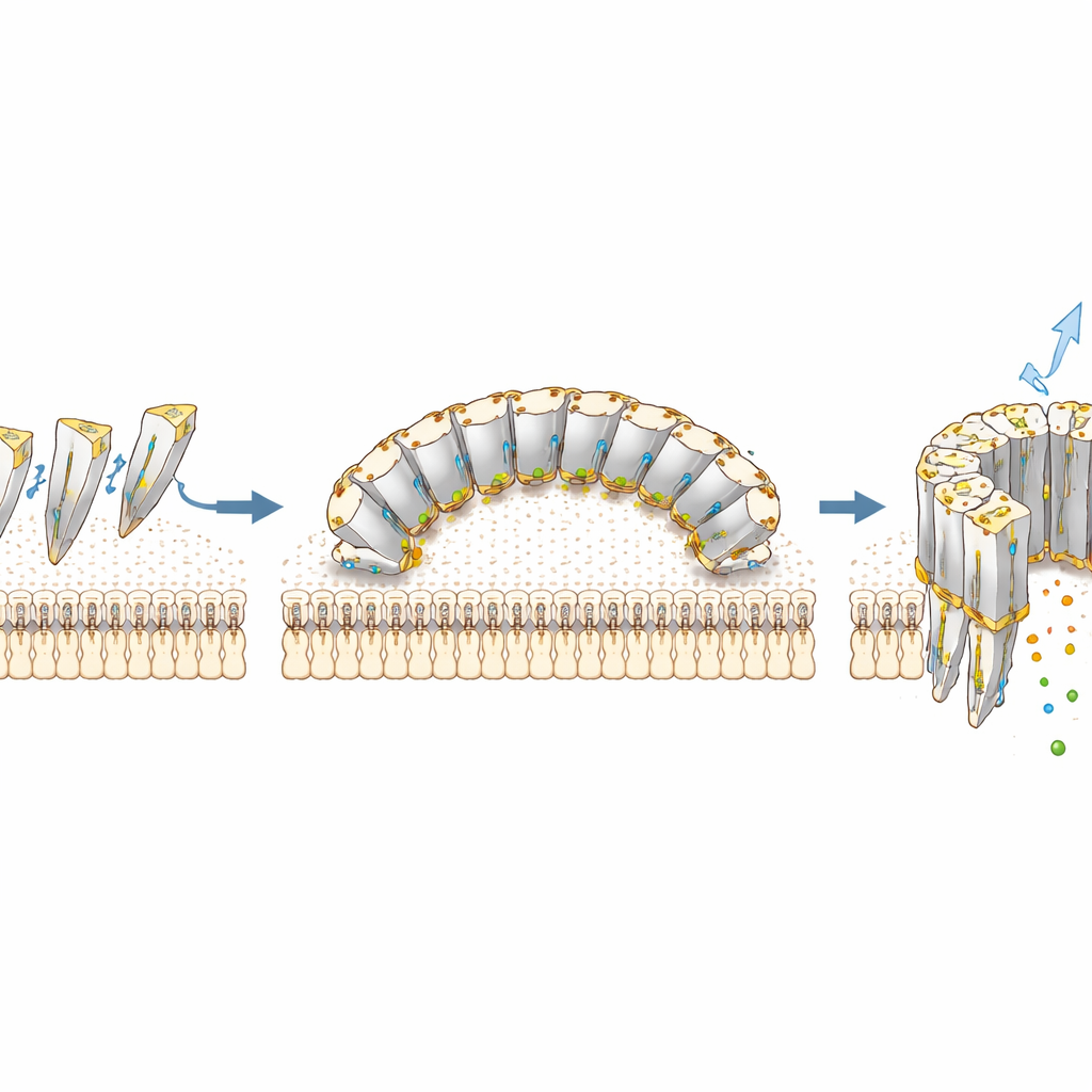
How GSDME Grabs the Membrane and Builds a Ring
The study pinpoints exactly how GSDME latches onto membranes and then links copies of itself into a complete ring. Two main contact regions act like grappling hooks: one, rich in positively charged building blocks, reaches toward negatively charged lipid headgroups, while a nearby cluster of greasy side chains dives into the fatty core of the membrane like an anchor. Mutating these residues in the lab almost completely shut down the protein’s ability to punch holes in artificial membranes and to kill cells, confirming their importance. Neighboring GSDME units then clasp each other through two separate contact surfaces, forming a sturdy chain that curves around into a full circle. Certain cancer-associated mutations cluster near these joining surfaces, suggesting they may blunt GSDME’s tumor-suppressing abilities by sabotaging pore assembly.
Switching GSDME On and Tuning Its Strength
Before GSDME can form pores, it must be cut in two by an internal executioner enzyme called caspase-3, which is best known for driving classical programmed cell death. The authors show that caspase-3 recognizes and cuts GSDME almost entirely based on a short, four–amino acid motif (DMPD) in a flexible linker between its halves, without needing to hold on to the protein’s tail region. This is different from another gasdermin, GSDMD, which requires extra docking contacts. As a result, GSDME can be activated rapidly wherever caspase-3 is triggered, such as in chemotherapy-treated tumor cells, redirecting them from a quiet death to an inflammatory one. After cleavage, GSDME’s activity is further boosted by the attachment of small fatty chains, a modification called palmitoylation, at specific cysteine sites—especially one buried in the membrane-spanning region. These fatty additions help the active fragment gather at membranes and assemble efficient pores, fine-tuning how violently a cell undergoes pyroptosis.
Why This Matters for Cancer and Inflammatory Disease
Together, these discoveries provide a structural and regulatory blueprint for how GSDME makes and controls membrane pores. For non-experts, the key idea is that cells use a two-step safety system: first, a cut by caspase-3 releases GSDME’s pore-forming half, and second, strategic fatty decorations make those pores more potent. By seeing exactly how GSDME grips membranes, links into rings, and is chemically tuned, researchers gain new entry points for drug design. Future therapies might enhance GSDME activity to help the immune system recognize and destroy tumors, or dampen it to protect tissues in inflammatory diseases where excessive cell explosion is harmful.
Citation: Teran, E., Tian, T., Wang, C. et al. Structural basis and regulation of GSDME pore formation. Nat Commun 17, 4148 (2026). https://doi.org/10.1038/s41467-026-70643-5
Keywords: pyroptosis, gasdermin E, caspase-3, palmitoylation, cancer immunology