Clear Sky Science · en
Functional and structural basis of a negative allostery within GABAB hetero-tetramers
Why brain signal brakes matter
Our brains rely on a delicate balance between “go” and “stop” signals. One of the most important brakes is the GABAB receptor, a protein on nerve cells that dampens activity and is targeted by medicines such as baclofen for muscle spasms and drugs used in addiction treatment. This study uncovers how groups of these receptors assemble and subtly restrain each other, revealing a built-in safety feature that can go wrong in neurological disease and that could be harnessed for better therapies.
Building blocks of a neural brake
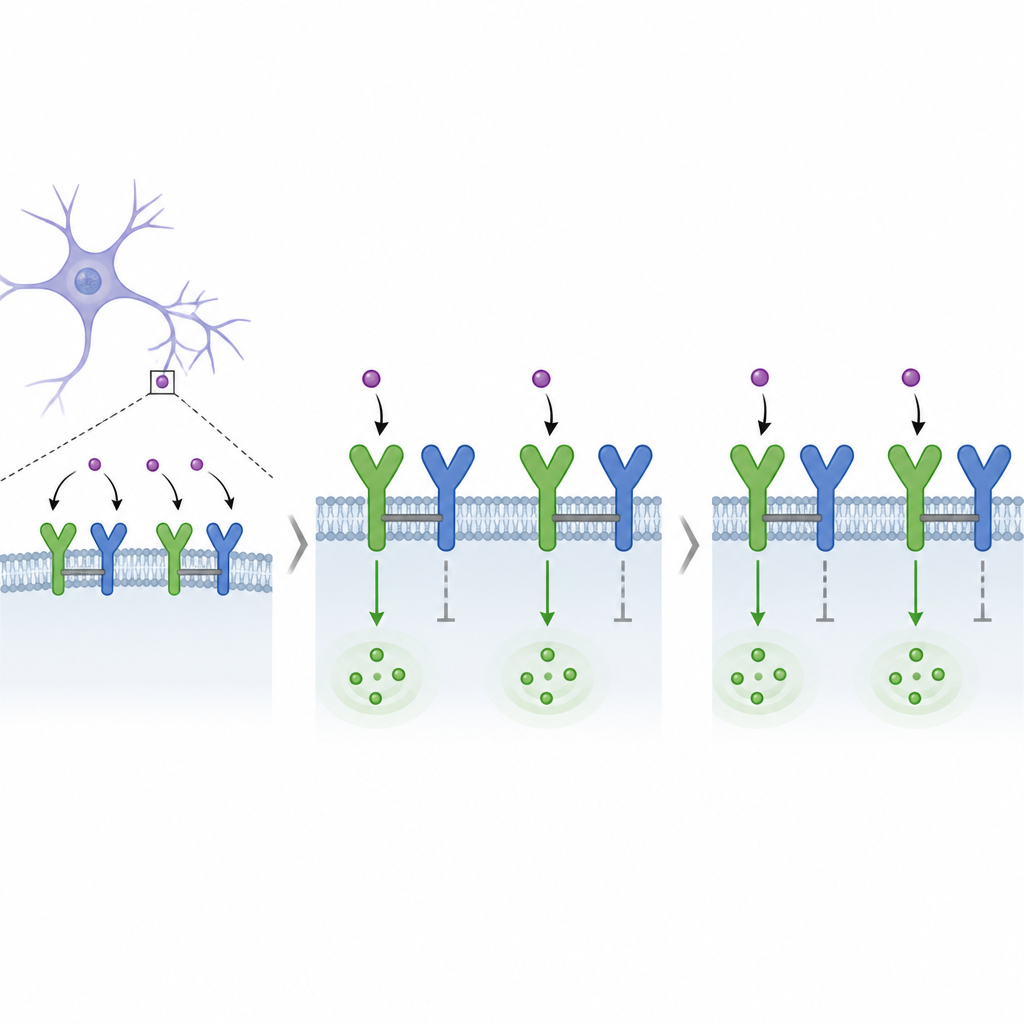
GABAB receptors sit on the surface of neurons and sense GABA, the brain’s main calming chemical. Each working receptor is made from two different protein subunits that come together as a pair: one side binds GABA, the other side talks to inside-the-cell signaling partners called G proteins. The authors revisit a long-standing question: do these pairs further group into larger clusters, and if so, do those clusters change how signals flow? Understanding this organization is crucial, because GABAB receptors influence movement, learning, memory, and mood, and are involved in conditions like epilepsy, anxiety, and pain.
Finding hidden receptor clusters in brain cells
To see how GABAB receptors are arranged in living cells, the team created tiny antibody fragments called nanobodies that latch very specifically onto each subunit. Using these as molecular tags in a microscopy method that lights up only when two tagged proteins are very close, they showed that pairs of GABAB receptors often sit side by side to form four-part complexes, called hetero-tetramers, in both engineered human cells and mouse neurons. They then used a light-emitting assay that reports when G proteins are recruited, allowing them to compare the signaling of simple two-part receptors with that of the four-part assemblies at the cell surface.
When one partner speaks, the other stays quiet
The researchers found that GABAB tetramers behave differently from single receptor pairs. Both the natural messenger GABA and a drug that binds deep within the receptor’s core could activate signaling, but within a tetramer only one pair appeared to signal efficiently at a time. Genetic mutations in the GABAB binding site that are linked to Rett-like syndromes and epileptic disorders mostly weakened signaling in tetramers, while leaving single pairs much less affected. This suggests that disease-causing changes in people may act largely by disturbing the special behavior of these larger assemblies rather than simply turning individual receptors on or off.
A crooked embrace that enforces silence
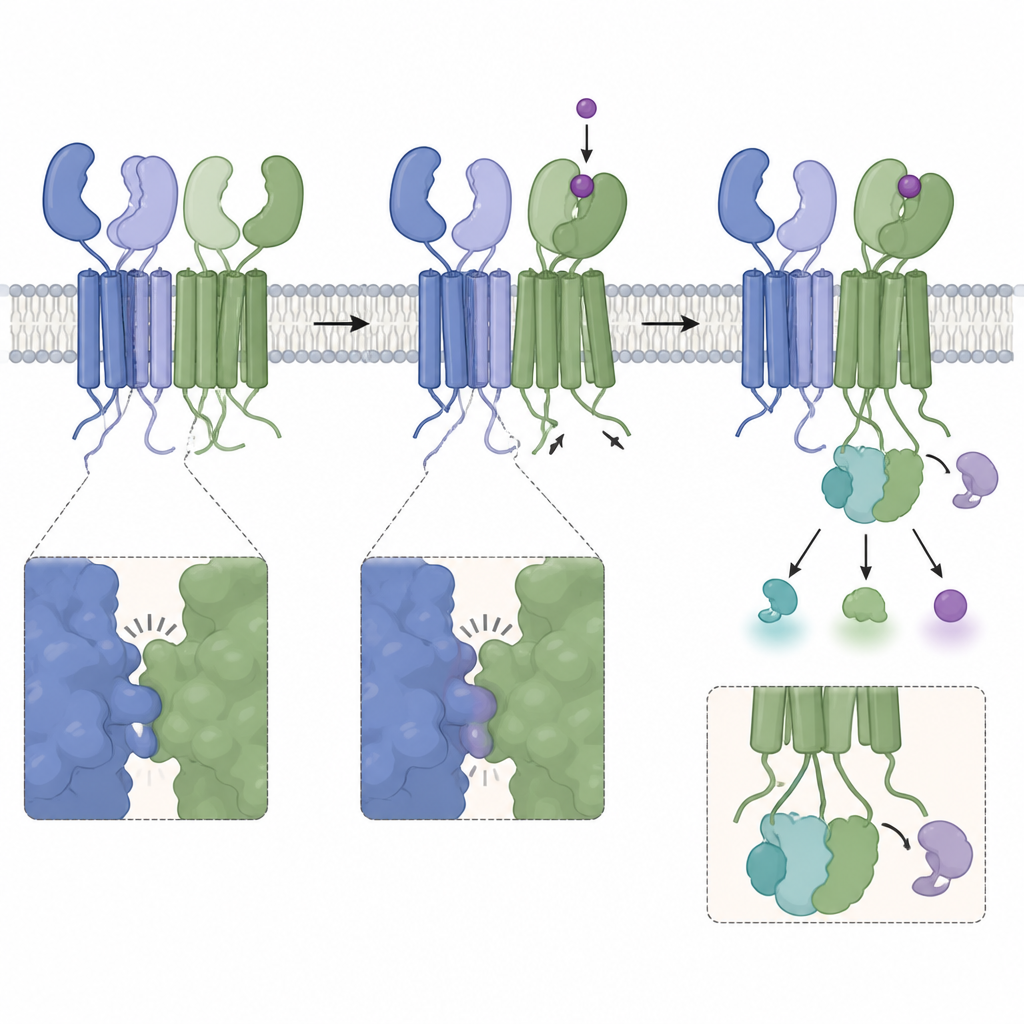
To understand how this selective silencing works, the team used cryo-electron microscopy to capture a high-resolution 3D picture of the human GABAB tetramer. The structure shows two receptor pairs joined through an uneven contact between their outer domains, where the GABA-binding regions of the two subunits meet in a lopsided fashion. Modeling indicates that if one binding pocket closes around a molecule, there is not enough room for the partner pocket to close at the same time. Mutations that insert a bulky sugar group or add extra amino acids at this interface loosen the contact and boost signaling, supporting the idea that the crooked embrace normally prevents both halves from activating together.
Linked cores that lock in the arrangement
Deeper in the membrane, the four buried segments of the tetramer form a more symmetrical diamond-like pattern. Each basic pair holds the same contact seen in inactive receptors, while additional contacts join one subunit from one pair to its neighbor from the other pair. Biochemical crosslinking experiments confirm that these membrane-spanning helices sit close enough to bond under the right conditions, explaining why tetramers are unusually stable once formed. Despite these tight links, drugs that act from within the membrane-spanning core still drive G protein activation from either pair, further highlighting that the main brake on signaling lies in the outer, GABA-binding regions.
What this means for brain health and future drugs
Overall, the study reveals that many GABAB receptors in neurons work in linked pairs of pairs, where an uneven contact between outer domains enforces a “one-at-a-time” rule for activation. This negative allostery, in which activation of one receptor pair suppresses its neighbor, provides a built-in way to limit the strength of inhibitory signals. Because several human mutations selectively damage the tetramer’s function, these complexes appear essential for normal brain activity. Appreciating this layered organization could guide the design of future drugs that either target the tetramers directly or fine-tune their internal tug-of-war, offering more precise ways to modulate the brain’s inhibitory brakes.
Citation: Shen, C., Ding, H., Zhang, S. et al. Functional and structural basis of a negative allostery within GABAB hetero-tetramers. Nat Commun 17, 4284 (2026). https://doi.org/10.1038/s41467-026-70640-8
Keywords: GABAB receptor, GPCR oligomerization, negative allostery, neural inhibition, cryo EM structure