Clear Sky Science · en
A conserved, immune-regulated peritrophin promotes Vibrio cholerae colonization of the arthropod intestine
A tiny helper with a big impact
Cholera is usually framed as a human disease spread by contaminated water, but the bacterium that causes it, Vibrio cholerae, spends much of its life interacting with small animals like insects and crustaceans. This study reveals how the microbe exploits a seemingly protective layer in these animals’ guts to settle in and thrive. By uncovering a conserved gut protein that actually helps V. cholerae stick to the intestinal lining, the work offers fresh insight into how environmental reservoirs of cholera are maintained—and hints at new ways to disrupt them.
The journey from water to insect gut
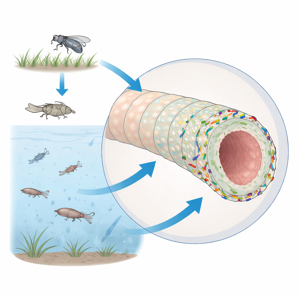
V. cholerae lives not only in human intestines but also in coastal waters, where it associates with a range of tiny arthropods such as copepods, rotifers, and midges. To model these environmental interactions, the researchers turned to the fruit fly, Drosophila melanogaster, a well-established system for studying gut biology and immunity. The fly midgut, roughly comparable to the human small intestine, is lined by an inner sheet of cells and covered toward the inside by a delicate, porous sleeve called the peritrophic matrix. This matrix is made of chitin—the same material found in insect shells—and chitin-binding proteins known as peritrophins. It acts like a semi-permeable raincoat: it lets nutrients pass but keeps many microbes at a distance from the gut cells.
When defense signals help the invader
Fruit fly gut cells mount rapid responses when they sense microbes. One key pathway, called IMD, detects bacterial molecules and turns on genes for antimicrobial peptides that can kill or restrain invading bacteria. A special group of hormone-producing cells in the front part of the midgut, the enteroendocrine cells, also use this pathway. In earlier work, these cells were shown to respond to bacterial byproducts by producing a small signaling peptide called tachykinin (Tk), which in turn influences metabolism and antimicrobial activity. The authors expected that turning down this immune signaling would give V. cholerae an easier time colonizing the gut. Instead, they found the opposite: blocking IMD or Tk specifically in enteroendocrine cells reduced bacterial colonization, whereas blocking the same pathway in nutrient-absorbing cells increased it. This pointed to a surprising conclusion: some products induced by Tk in hormone-producing cells actually favor V. cholerae’s ability to stick around.
A sticky protein on the gut’s inner shield
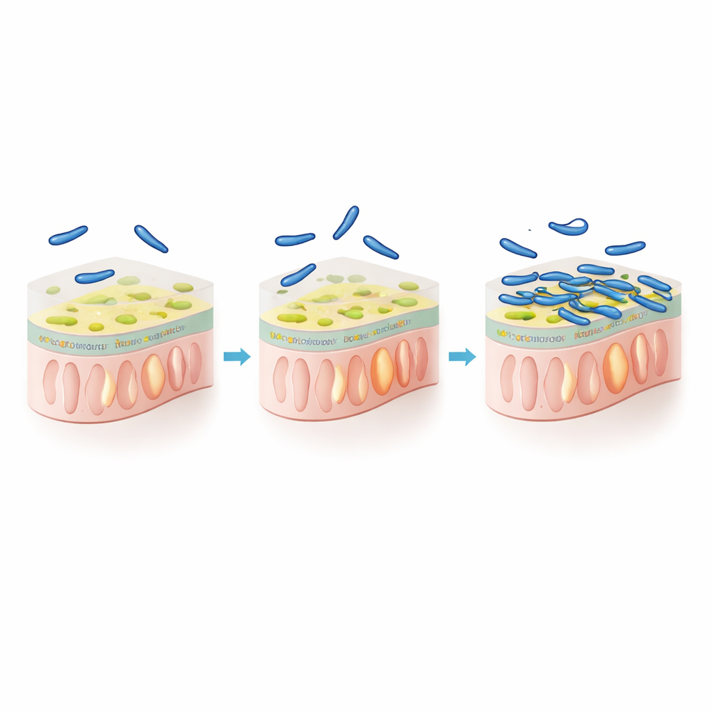
To identify those helpful host factors, the team compared gene activity in normal flies and flies where Tk was silenced in enteroendocrine cells. Among the genes that dropped when Tk was reduced were several involved in chitin handling, including a small secreted protein called Peritrophin-15a (Peri-15a). Peri-15a is produced mainly by enteroendocrine cells in the front midgut and is built to bind chitin, positioning it in the peritrophic matrix. When the scientists knocked down Peri-15a in all hormone-producing gut cells, V. cholerae colonization fell by roughly a hundredfold, even though standard immune markers and Tk levels barely changed. Infection with V. cholerae itself, or feeding flies a steroid hormone known to boost gut immunity, both drove up Peri-15a levels and, correspondingly, bacterial colonization—but this boost vanished when Peri-15a was suppressed. Importantly, detailed imaging experiments showed that removing Peri-15a did not make the matrix leaky or structurally fragile, suggesting its key role is not to maintain the barrier, but to offer a better foothold for the bacteria.
Balancing digestion, protection, and attachment
The peritrophic matrix must remain porous enough for food breakdown while shielding the gut from constant immune irritation. The authors probed how this balance interacts with V. cholerae attachment by altering chitin-degrading enzymes in the fly and the bacterium. Reducing a major fly chitinase made the matrix denser, dampened immune signaling, lowered Peri-15a levels, and reduced bacterial colonization. In contrast, disabling a master regulator of V. cholerae’s own chitin digestion machinery modestly increased colonization—an effect that disappeared when Tk or Peri-15a were knocked down. These patterns support a model in which Peri-15a decorates the matrix without greatly changing its porosity, while V. cholerae benefits most when its binding sites on this protein-rich surface are preserved rather than chewed away by its own enzymes.
A shared strategy across water worlds
Searching structural databases, the researchers found close Peri-15a look-alikes in a wide range of insects and in marine zooplankton known to harbor Vibrio species. Prior work in copepods has shown that Vibrio colonization boosts expression of similar chitin-binding proteins. Taken together, these observations suggest that V. cholerae has tapped into a common feature of arthropod gut biology: an immune-regulated, chitin-binding coat that can double as a docking surface. For a lay audience, the key message is that the same immune signals that help small aquatic animals defend and maintain their guts can inadvertently create sticky landing pads that stabilize cholera bacteria in the wild. Understanding this finely tuned interaction between host shield and microbe hitchhiker could inform new strategies to break environmental links in the cholera transmission chain.
Citation: Barraza, D., Paulo, T.F., Findley, L. et al. A conserved, immune-regulated peritrophin promotes Vibrio cholerae colonization of the arthropod intestine. Nat Commun 17, 3920 (2026). https://doi.org/10.1038/s41467-026-70629-3
Keywords: Vibrio cholerae, arthropod gut, peritrophic matrix, chitin-binding proteins, environmental reservoirs