Clear Sky Science · en
FineST: contrastive learning integrates histology and spatial transcriptomics for nuclei-resolved ligand-receptor analysis
Seeing Hidden Conversations Between Cells
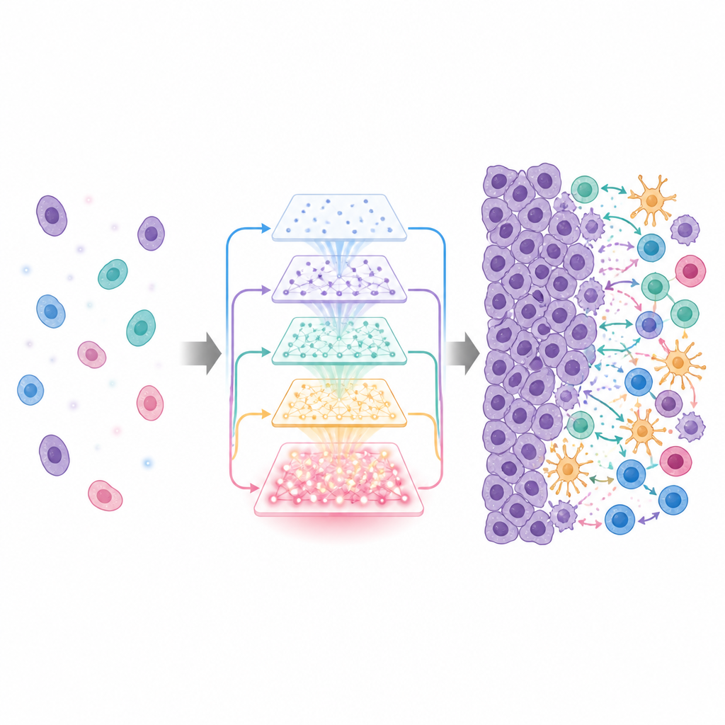
Our bodies are full of tiny conversations. Neighboring cells constantly send and receive chemical messages that help tissues grow, fight infections, or, in the case of cancer, outsmart the immune system. This study introduces FineST, a new computational tool that combines microscope images and gene activity maps to reveal these subtle cell-to-cell talks at the level of individual cell nuclei, offering a sharper view of how tumors interact with their surroundings.
Why Spatial Maps of Cells Fall Short
Modern techniques in spatial transcriptomics can record which genes are active and where in a tissue slice. However, many popular platforms capture signals from several cells at once in each spot, and much of the tissue has missing or extremely sparse measurements. This low resolution and patchy data make it hard to pinpoint which specific cells are talking to each other and through which molecular signals. As a result, many existing methods either ignore the physical distance between cells or cannot resolve communication at single-cell scale, especially for the critical ligand-receptor pairs that carry these messages.
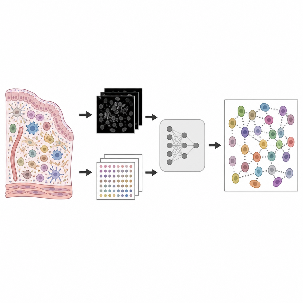
Fusing Microscopy and Gene Activity with FineST
FineST tackles this problem by tightly integrating high-resolution histology images with spatial gene expression data. It uses a powerful image-recognition model, originally trained on large collections of tissue slides, to break each coarse measurement spot into many tiny tiles roughly the size of a single cell. By learning how image patterns relate to the underlying gene activity, FineST can impute detailed RNA expression for each tile and even around the center of individual nuclei. A contrastive learning strategy aligns the image features and gene profiles in a shared space so that matching regions reinforce each other, while additional reconstruction steps keep the predicted RNA patterns faithful to the original data.
Sharper Views of Tumors and Their Neighborhoods
The researchers tested FineST on several cancer datasets, including colorectal, breast, liver, and nasopharyngeal tumors. In high-definition colorectal tissue, FineST more accurately recovered the spatial patterns of hundreds of signaling genes than a leading alternative method, and it better predicted the mix of cell types in each region. In breast cancer samples measured by two different platforms, FineST’s reconstructions at near single-nucleus resolution matched detailed ground truth maps and revealed subtle differences between pre-invasive and more invasive tumor areas. It highlighted specific cell populations, such as specialized myoepithelial cells, that appeared in one early lesion but were largely absent in more aggressive regions, hinting at changes linked to progression.
Tracing Tumor–Immune Interactions
FineST is especially powerful for mapping how tumors interact with immune and support cells. In nasopharyngeal carcinoma, it identified hundreds of ligand-receptor pairs that were spatially co-expressed when examined at nucleus-level resolution, far more than at the original coarse spot level. These patterns lined up with distinct regions rich in T cells, B cells, regulatory T cells, and tumor cells, and were enriched for pathways related to immune presentation, cell growth, and movement. Within small regions known as tertiary lymphoid structures, FineST uncovered concentrated communication between tumor cells, T cells, B cells, and fibroblasts, including inhibitory signals that may help the cancer evade immune attack.
Clues to Treatment Resistance
In liver cancer patients treated with PD-1 immune therapy, FineST helped clarify why some tumors resist treatment. By enhancing weak gene signals in the original data, it revealed more coherent patterns of macrophages and cancer-associated fibroblasts forming a barrier around the tumor, and pinpointed ligand-receptor interactions that were active only in non-responders. Some of these interactions, including those involving the PD-1 pathway and additional signaling routes, appeared precisely along the boundary between tumor and immune cells, suggesting how communication across this barrier might suppress immune attack.
What This Means Going Forward
To a non-specialist, FineST can be viewed as a microscope upgrade in software: it uses existing images and gene measurements to reconstruct who is talking to whom inside a tissue, cell by cell. By resolving these conversations at the level of individual nuclei and linking them to known biological pathways, the method can expose how tumor cells influence neighboring immune and support cells, how certain regions become more invasive, and why some patients fail to respond to immunotherapy. As spatial technologies continue to improve, tools like FineST are poised to turn complex tissue snapshots into clear, interpretable maps of cellular dialogue.
Citation: Li, L., Wang, T., Liang, Z. et al. FineST: contrastive learning integrates histology and spatial transcriptomics for nuclei-resolved ligand-receptor analysis. Nat Commun 17, 4645 (2026). https://doi.org/10.1038/s41467-026-70528-7
Keywords: spatial transcriptomics, cell-cell communication, cancer microenvironment, histology integration, ligand-receptor analysis