Clear Sky Science · en
Megakaryocyte and platelet thrombospondin-1 regulates matrix remodeling by stabilizing basement membrane COL6A1 in lung injury
Why protecting the lung’s scaffolding matters
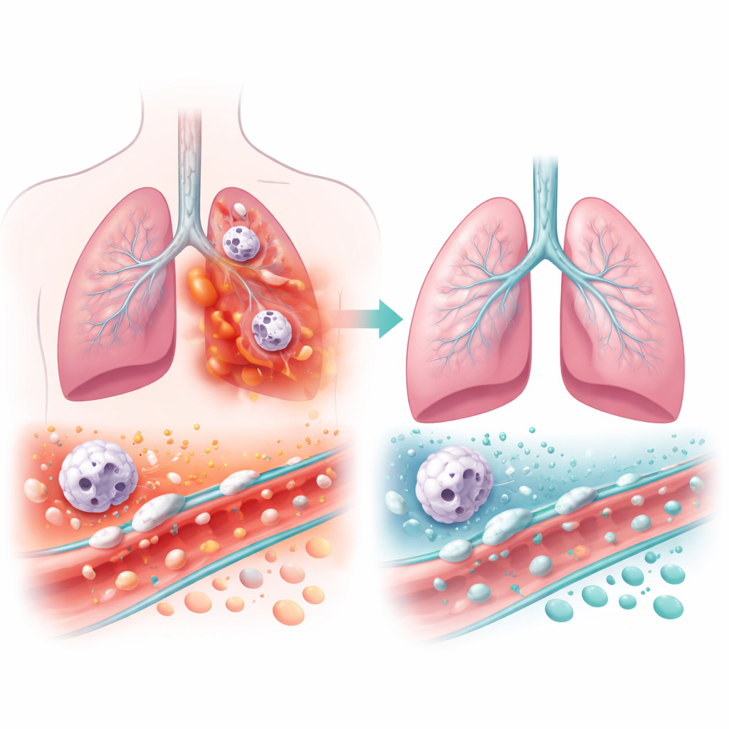
When the lungs are injured by severe infections or other insults, people can develop a life‑threatening condition called acute respiratory distress syndrome, where the tiny air sacs flood and breathing fails. Doctors know that inflammation and scar‑forming tissue changes are central to this process, but less is known about how the body’s own blood cells try to protect the lung’s delicate support structure during the earliest hours of damage. This study uncovers an unexpected defender: a protein released by specialized bone marrow cells and platelets that helps hold the lung’s microscopic scaffolding together and keep harmful white blood cells in check.
A closer look at the lung’s hidden framework
Deep inside the lung, each air sac is wrapped in a thin sheet of proteins called the basement membrane. This layer acts like reinforcing mesh in concrete, giving strength to the tissue while allowing oxygen to pass into nearby blood vessels. In acute lung injury and its severe form, acute respiratory distress syndrome, this framework is rapidly remodeled: sturdy fibers are broken down and replaced by looser, more fibrous material that stiffens the lung and lets fluid leak into the air spaces. The authors focused on a basement membrane protein called COL6A1 and on thrombospondin‑1, a sticky “matricellular” protein that can bind many matrix components and is stored in large amounts inside platelets and their parent cells, megakaryocytes.
Platelet protein as an early guardian of the barrier
Using genetically engineered mice and advanced imaging that tracks events in living lungs in real time, the team deleted thrombospondin‑1 specifically from megakaryocytes and platelets. These mice had normal platelet counts and clotting responses, showing that basic blood‑clotting function was intact. But when the lungs were injured by bacterial products or live Pseudomonas aeruginosa infection, the picture changed dramatically: fluorescent dyes leaked much more readily from blood vessels into the air sacs, microscopic lung injury scores rose, and lung mechanics worsened, indicating stiffer, less compliant lungs. Despite similar numbers of neutrophils—the first‑responder white blood cells—in the airspaces, these cells were far more activated, releasing higher levels of destructive enzymes such as neutrophil elastase and MMP‑9.
How missing protection reshapes the lung matrix
To understand what was happening to the tissue scaffold itself, the researchers combined laser‑guided sampling of injured alveolar regions with high‑resolution mass spectrometry. They found that lungs lacking megakaryocyte and platelet thrombospondin‑1 accumulated markers of a “provisional” repair matrix, including fibrin and fibronectin, along with elevated neutrophil proteases. At the same time, key basement membrane components, especially COL6A1, were depleted. Imaging methods that visualize collagen fibers without dyes revealed thicker fibrillar collagen deposits, particularly around areas where fluorescent leak and large megakaryocyte cells clustered. In other words, without this platelet‑derived protein, the fine basement membrane mesh was eroded and replaced by coarser collagen fibers, altering both barrier integrity and local cell behavior.
Neutrophils, megakaryocytes, and a feedback loop of damage
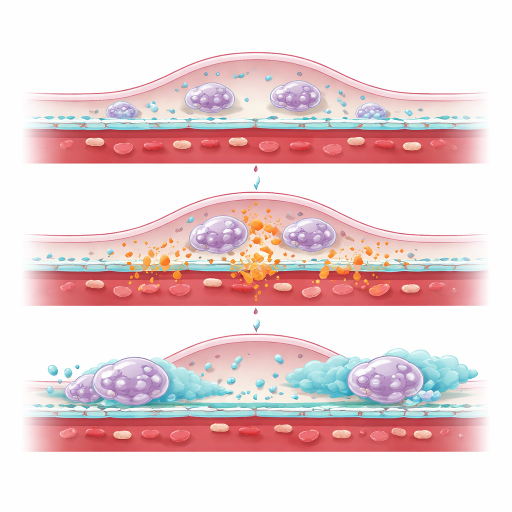
The study also shows that thrombospondin‑1 helps restrain a damaging feedback loop between neutrophils and megakaryocytes. In mice missing the protein in these blood cells, megakaryocytes accumulated in injured lungs near exposed collagen and leak sites, while circulating platelet numbers fell. When neutrophils were selectively depleted with antibodies, lung leak dropped toward normal, megakaryocyte numbers in the lung declined, and levels of basement membrane proteins COL4A1 and COL6A1 in lung fluid rose. Pro‑collagen type I, a marker of new fibrillar collagen production likely coming from fibroblasts, remained elevated, suggesting that many cell types participate in rebuilding the scaffold, but thrombospondin‑1 is particularly important for shielding the basement membrane from neutrophil‑driven enzymatic attack.
What this means for patients with severe lung injury
Together, these findings reveal that megakaryocytes and platelets do more than plug bleeding vessels: by releasing thrombospondin‑1 at sites of lung injury, they help preserve the thin protein sheet that underpins each air sac and temper the destructive tendencies of recruited neutrophils. When this protective system is missing, the basement membrane protein COL6A1 is lost, leaks worsen, and the lung rapidly fills with a less organized collagen network that stiffens the tissue. For people with conditions like severe pneumonia or acute respiratory distress syndrome, therapies that boost or mimic this platelet‑derived protection could one day offer a way to limit early barrier failure and guide the lung toward more effective repair rather than scarring.
Citation: Peñaloza, H.F., Gheware, A., Gupta, A. et al. Megakaryocyte and platelet thrombospondin-1 regulates matrix remodeling by stabilizing basement membrane COL6A1 in lung injury. Nat Commun 17, 3844 (2026). https://doi.org/10.1038/s41467-026-70489-x
Keywords: acute lung injury, platelets, neutrophils, extracellular matrix, thrombospondin-1