Clear Sky Science · en
Cryo-ET comparison of the hierarchical ultrastructure of silkworm, spider, and artificial silk fibers
Why silk is more than just a pretty thread
Silk is famous for its shine and softness, but its real superpower is hidden deep inside each strand. Silkworm and spider silks can be stronger than steel of the same thickness and far tougher than many modern plastics. Scientists and engineers dream of copying this natural fiber to make better medical implants, smart clothing, and eco-friendly materials. This study peels back silk’s structure layer by layer, comparing natural silks from silkworms and spiders with man‑made silk, to find out what makes the real thing so special—and why our artificial versions still fall short. 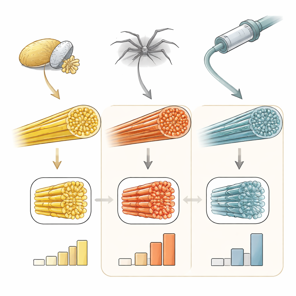
Looking inside silk without disturbing it
To see how silk is built from the inside out, the researchers used a powerful imaging approach borrowed from cell biology. First, they sliced frozen silk fibers into ultra‑thin sheets using a focused beam of ions, a technique called cryo‑FIB milling. Then they imaged these sheets in three dimensions with cryo‑electron tomography, which records many tilted views and reconstructs a 3D picture. Because the fibers are flash‑frozen in a watery state, this method avoids the harsh chemicals and stains that can squash or clump delicate structures. The result is an unusually faithful view of how the proteins that make up silk are actually arranged inside intact fibers.
The tiniest building blocks: beaded nano‑threads
Silk is largely made from a protein called fibroin in silkworms and spidroin in spiders. For years, scientists debated whether these proteins travel and assemble as spheres, rods, or some other shape. By looking at material taken from silkworm silk glands, the team found that fibroin forms very thin, flexible threads only about 3.6 to 4 nanometers thick—tens of thousands of times thinner than a human hair. Each of these “nanofibrils” looks like a string of tiny beads, rather than a smooth rod, suggesting that individual parts of the protein fold into small globular segments along a flexible chain. These same nanofibrils, with nearly identical size and appearance, were also found inside fully formed silkworm silk fibers, showing that the basic building blocks survive the journey from liquid dope in the gland to solid thread emerging from the spinneret.
How natural and artificial silks pack their nano‑threads
Inside the silkworm fiber, the nanofibrils run mostly parallel to the length of the thread but do not pack perfectly. Regions of tight bundling are interspersed with looser zones and visible gaps, and neighboring nanofibrils are often linked by short cross‑bridges. When the researchers examined spider dragline silk, they saw a striking contrast: the same kind of nanofibrils were packed far more densely and almost flawlessly aligned with the fiber axis, leaving almost no empty space. This dense, orderly packing likely underlies the spider fiber’s superior strength and toughness. In man‑made silk spun from regenerated silkworm protein, however, the internal picture was very different. The nanofibrils showed poor alignment, uneven density, and a generally disordered layout, suggesting that current spinning conditions do not reproduce the carefully controlled environment of the animal’s silk gland. 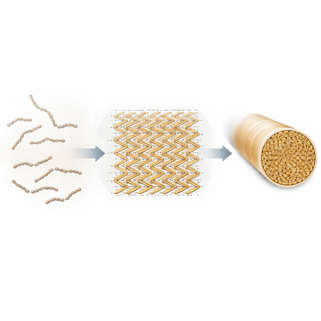
A hidden pattern that shapes the whole fiber
Looking more closely at silkworm silk, the team discovered that the nanofibrils do not simply lie side‑by‑side: they form a repeating herringbone‑like pattern. In this pattern, rows of fibrils tilt in alternating directions, producing an anisotropic, or directionally biased, arrangement. Multiple layers of this herringbone pattern then stack up to build the full‑sized fiber. Importantly, this same pattern had previously been seen near the spinneret inside the silk gland, and the new work shows it persists all the way into the final thread. The pattern appears to be organized around invisible axes and may involve helper molecules beyond fibroin itself. A similar motif was even observed in parts of the spider’s spinning duct, hinting that this higher‑order layout is a shared solution evolved in different silk‑producing animals to balance strength, toughness, and flexibility.
What this means for future super‑silks
By revealing that both silkworm and spider silks are built from extremely thin, beaded nanofibrils arranged in layered herringbone patterns—and by showing how tightly and neatly those nanofibrils pack in natural fibers compared with artificial ones—this study provides a structural roadmap for designing better synthetic silks. The work suggests that simply copying the protein recipe is not enough; the liquid environment, flow, and alignment during spinning must also be carefully controlled to reproduce nature’s remarkable architecture. Understanding how these tiny nano‑threads organize themselves into robust fibers could guide the creation of next‑generation materials that are strong, tough, lightweight, and biodegradable.
Citation: Song, K., Zhang, H., Zhang, X. et al. Cryo-ET comparison of the hierarchical ultrastructure of silkworm, spider, and artificial silk fibers. Nat Commun 17, 3608 (2026). https://doi.org/10.1038/s41467-026-70477-1
Keywords: silk nanofibrils, spider silk, silkworm silk, cryo electron tomography, artificial silk fibers