Clear Sky Science · en
Brachiopod genome unveils the evolution of BMP signalling in bilaterian body patterning
How early embryos decide top from bottom
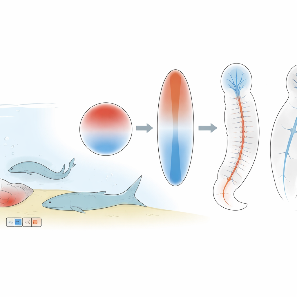
Every animal body, from worms to humans, has a built-in “top–bottom” (back–belly) axis that forms very early in the embryo. This paper explores how a little-known marine animal, the brachiopod Lingula anatina, uses a chemical signalling system to set up that axis and decide where its nervous system should form. By comparing brachiopods with other animals, the authors uncover ancient rules that appear to have guided body patterning across most of the animal kingdom.
A shell-bearing worm cousin as a living fossil
Brachiopods look a bit like clams, with two shells, but they belong to their own branch of animal life and have a long fossil history. They are part of the spiralians, a huge group that also includes molluscs and segmented worms, well known for diverse and sometimes puzzling ways of building their body axes. To understand how brachiopods fit into this picture, the researchers first produced a high-quality, chromosome-level genome for Lingula anatina. This allowed them to catalogue all the key genes involved in one crucial signalling system, known as the BMP pathway, which helps cells know whether they will become back, belly, skin, or nerve tissue. They found that Lingula carries a mostly “textbook” set of these genes, in simple single copies, making it an excellent reference for comparing with other spiralians.
Ancient body-plan genes locked into place
Looking across genomes from several animal groups, the team discovered that BMP-related genes are not only conserved in sequence but also in their broad chromosomal neighborhoods. Even though chromosomes have been shuffled and fused differently in worms, molluscs, brachiopods, and early chordates, the BMP pathway genes tend to stay on the same ancestral chromosome blocks. Similar stability was seen for other body-plan genes such as Wnt and Hox. This suggests that, over hundreds of millions of years, evolution has strongly resisted moving these developmental control genes away from their surrounding regulatory regions, preserving a hidden genomic “map” for building body axes.
A chemical seesaw that defines back and belly
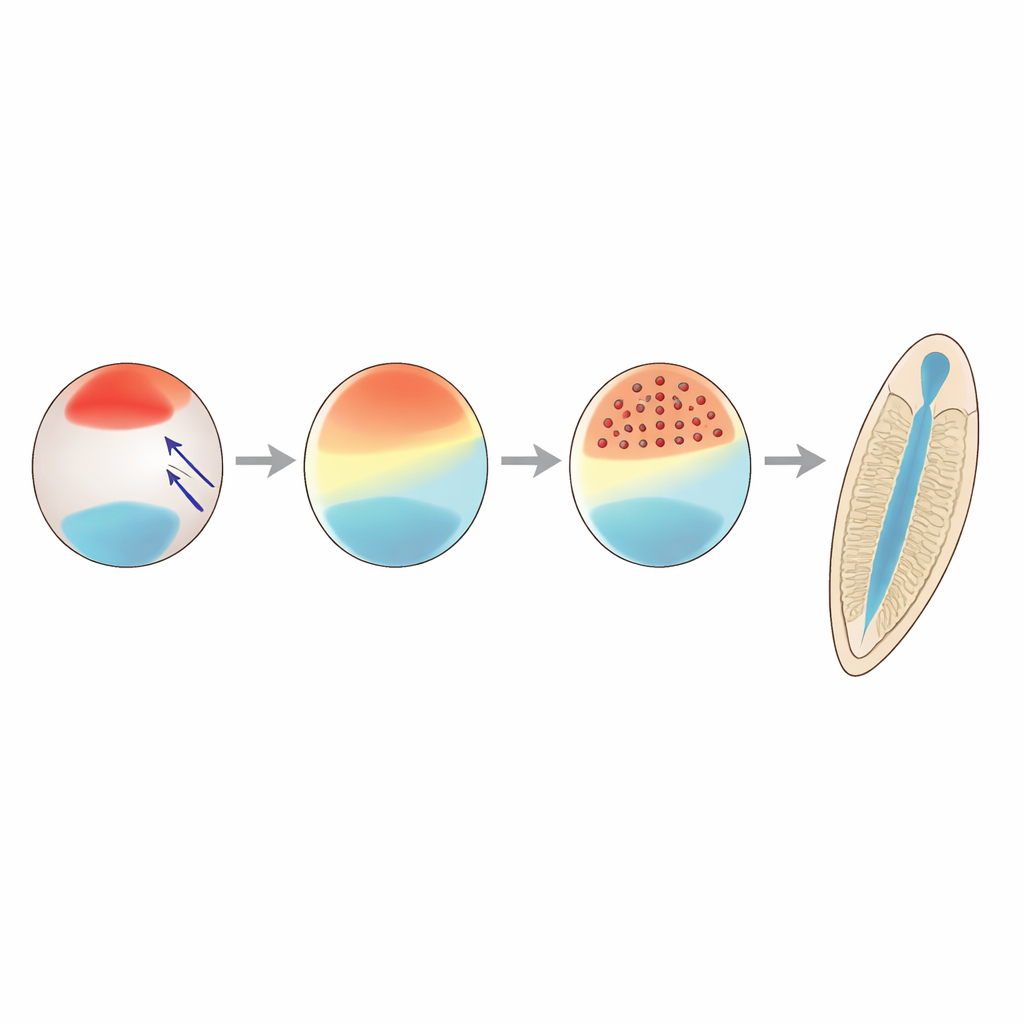
To see how the BMP system actually works in Lingula embryos, the authors measured when and where BMP genes and their antagonists are active, and tracked the signal inside cells using antibodies. They found a striking “seesaw” arrangement: different BMP ligands are produced on opposite sides of the embryo, while an inhibitory molecule, chordin, is concentrated on the future belly side. Together these sources create a gradient of BMP activity that is high on the future back and low on the belly. This asymmetry first appears around the blastula stage and persists through gastrulation, matching patterns seen in insects and some marine deuterostomes. When the researchers blocked BMP receptors, the gradient disappeared and gastrulation faltered; when they added extra BMP protein, signalling spread over the whole embryo and normal shaping was disturbed, including the separation of the two larval shell lobes.
Keeping nerves away from the high-signal side
The team then asked how this gradient influences nervous system formation. Using RNA sequencing, they compared normal embryos with ones in which BMP signalling was either blocked or overactivated. Dozens of genes typically associated with nerve cells and brain development in other animals were strongly suppressed when BMP was high and expanded when BMP was low. In situ hybridisation showed that key neural markers sit opposite the BMP maximum, on the low-signal belly side where chordin is expressed. When BMP was inhibited, these neural domains spread; when BMP was boosted, several neural markers vanished or shrank. At the same time, many genes involved in DNA replication and cell division were turned down by high BMP, fitting with observed reductions in cell proliferation.
Shared rules across distant animal branches
Comparing their results with the classic frog model Xenopus, the authors found not only similar responses of neural genes to BMP, but also parallel regulation of several organiser genes that shape the dorsal–ventral axis. Despite differences in embryo geometry and an evolutionary “flip” of the axis in chordates, the core logic is the same: a BMP–chordin system creates a gradient, and neural tissue arises where BMP activity is low. The authors argue that this arrangement—and even the BMP “seesaw” of ligands on opposite sides—likely existed in the last common ancestor of bilaterian animals. Over time, many spiralian lineages appear to have tinkered with the downstream wiring of this system, leading to today’s diversity in early development, but the central gradient remains a deeply conserved scaffold.
What this means for understanding body plans
For a non-specialist, the take-home message is that very different animals, from shell-bearing brachiopods to frogs and flies, seem to share an ancient chemical strategy for deciding which side becomes back or belly and where the nervous system should form. Lingula shows that spiralians, despite their varied embryonic tricks, retain this core BMP-based patterning system. Evolution has experimented mainly with the “downstream” details, not the overall blueprint. By combining a new reference genome with precise experiments in living embryos, this study helps reveal how a simple gradient of a few signalling molecules can underlie the rich diversity of animal body plans we see today.
Citation: Lewin, T.D., Sakagami, T., Shimizu, K. et al. Brachiopod genome unveils the evolution of BMP signalling in bilaterian body patterning. Nat Commun 17, 3856 (2026). https://doi.org/10.1038/s41467-026-70403-5
Keywords: BMP signalling, dorsal–ventral patterning, brachiopod development, neural induction, evolutionary developmental biology